 Movie
Movie Controller

[English] 日本語
 Yorodumi
- PDB-8b9f: Structure of Echovirus 11 complexed with DAF (CD55) calculated fr... -
+ Open data

- Basic information
| Entry | Database: PDB / ID: 8b9f | ||||||
|---|---|---|---|---|---|---|---|
| Title | Structure of Echovirus 11 complexed with DAF (CD55) calculated from symmetry expansion | ||||||
 Components Components |
| ||||||
 Keywords Keywords | VIRUS / Echovirus / receptor / CD55 / DAF | ||||||
| Function / homology |  Function and homology information Function and homology informationregulation of lipopolysaccharide-mediated signaling pathway / negative regulation of complement activation / regulation of complement-dependent cytotoxicity / regulation of complement activation / respiratory burst / positive regulation of CD4-positive, alpha-beta T cell activation / positive regulation of CD4-positive, alpha-beta T cell proliferation / Class B/2 (Secretin family receptors) / symbiont-mediated suppression of host cytoplasmic pattern recognition receptor signaling pathway via inhibition of RIG-I activity / ficolin-1-rich granule membrane ...regulation of lipopolysaccharide-mediated signaling pathway / negative regulation of complement activation / regulation of complement-dependent cytotoxicity / regulation of complement activation / respiratory burst / positive regulation of CD4-positive, alpha-beta T cell activation / positive regulation of CD4-positive, alpha-beta T cell proliferation / Class B/2 (Secretin family receptors) / symbiont-mediated suppression of host cytoplasmic pattern recognition receptor signaling pathway via inhibition of RIG-I activity / ficolin-1-rich granule membrane / complement activation, classical pathway / transport vesicle / side of membrane / COPI-mediated anterograde transport / endoplasmic reticulum-Golgi intermediate compartment membrane / secretory granule membrane / Regulation of Complement cascade / picornain 2A / symbiont-mediated suppression of host mRNA export from nucleus / symbiont genome entry into host cell via pore formation in plasma membrane / picornain 3C / T=pseudo3 icosahedral viral capsid / ribonucleoside triphosphate phosphatase activity / positive regulation of T cell cytokine production / host cell cytoplasmic vesicle membrane / nucleoside-triphosphate phosphatase / channel activity / positive regulation of cytosolic calcium ion concentration / virus receptor activity / monoatomic ion transmembrane transport / DNA replication / RNA helicase activity / membrane raft / endocytosis involved in viral entry into host cell / Golgi membrane / symbiont-mediated activation of host autophagy / innate immune response / RNA-directed RNA polymerase / cysteine-type endopeptidase activity / viral RNA genome replication / RNA-directed RNA polymerase activity / DNA-templated transcription / Neutrophil degranulation / lipid binding / virion attachment to host cell / host cell nucleus / structural molecule activity / cell surface / proteolysis / RNA binding / extracellular exosome / extracellular region / zinc ion binding / ATP binding / membrane / plasma membrane Similarity search - Function | ||||||
| Biological species |  Homo sapiens (human) Homo sapiens (human) 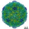 Echovirus E11 Echovirus E11 | ||||||
| Method | ELECTRON MICROSCOPY / single particle reconstruction / cryo EM / Resolution: 3.93 Å | ||||||
 Authors Authors | Stuart, D.I. / Ren, J. / Qin, L. / Zhou, D. | ||||||
| Funding support |  United Kingdom, 1items United Kingdom, 1items
| ||||||
 Citation Citation | Journal: Viruses / Year: 2022 Title: Switching of Receptor Binding Poses between Closely Related Enteroviruses. Authors: Daming Zhou / Ling Qin / Helen M E Duyvesteyn / Yuguang Zhao / Tzou-Yien Lin / Elizabeth E Fry / Jingshan Ren / Kuan-Ying A Huang / David I Stuart /  Abstract: Echoviruses, for which there are currently no approved vaccines or drugs, are responsible for a range of human diseases, for example echovirus 11 (E11) is a major cause of serious neonatal morbidity ...Echoviruses, for which there are currently no approved vaccines or drugs, are responsible for a range of human diseases, for example echovirus 11 (E11) is a major cause of serious neonatal morbidity and mortality. Decay-accelerating factor (DAF, also known as CD55) is an attachment receptor for E11. Here, we report the structure of the complex of E11 and the full-length ectodomain of DAF (short consensus repeats, SCRs, 1-4) at 3.1 Å determined by cryo-electron microscopy (cryo-EM). SCRs 3 and 4 of DAF interact with E11 at the southern rim of the canyon via the VP2 EF and VP3 BC loops. We also observe an unexpected interaction between the N-linked glycan (residue 95 of DAF) and the VP2 BC loop of E11. DAF is a receptor for at least 20 enteroviruses and we classify its binding patterns from reported DAF/virus complexes into two distinct positions and orientations, named as E6 and E11 poses. Whilst 60 DAF molecules can attach to the virion in the E6 pose, no more than 30 can attach to E11 due to steric restrictions. Analysis of the distinct modes of interaction and structure and sequence-based phylogenies suggests that the two modes evolved independently, with the E6 mode likely found earlier. | ||||||
| History |
|
- Structure visualization
| Structure viewer | Molecule: MolmilJmol/JSmol |
|---|
- Downloads & links
-Download
| PDBx/mmCIF format | 8b9f.cif.gz | 162.3 KB | Display | PDBx/mmCIF format |
|---|---|---|---|---|
| PDB format | pdb8b9f.ent.gz | 119.4 KB | Display | PDB format |
| PDBx/mmJSON format | 8b9f.json.gz | Tree view | PDBx/mmJSON format | |
| Others |  Other downloads Other downloads |
-Validation report
| Summary document | 8b9f_validation.pdf.gz | 1.4 MB | Display | wwPDB validaton report |
|---|---|---|---|---|
| Full document | 8b9f_full_validation.pdf.gz | 1.4 MB | Display | |
| Data in XML | 8b9f_validation.xml.gz | 35.6 KB | Display | |
| Data in CIF | 8b9f_validation.cif.gz | 50.2 KB | Display | |
| Arichive directory | https://data.pdbj.org/pub/pdb/validation_reports/b9/8b9fftp://data.pdbj.org/pub/pdb/validation_reports/b9/8b9f | HTTPS FTP |
-Related structure data
| Related structure data |  15930MC  8b8rC M: map data used to model this data C: citing same article ( |
|---|---|
| Similar structure data |
-Links
 PDBj
PDBj


- Assembly
| Deposited unit | 
|
|---|---|
| 1 |
|
-Components
| #1: Protein | Mass: 29771.059 Da / Num. of mol.: 1 Source method: isolated from a genetically manipulated source Source: (gene. exp.) Homo sapiens (human) / Gene: CD55, CR, DAF / Cell line (production host): HEK293T / Production host: Homo sapiens (human) / References: UniProt: P08174 |
|---|---|
| #2: Protein | Mass: 28886.428 Da / Num. of mol.: 1 Source method: isolated from a genetically manipulated source Source: (gene. exp.) Echovirus E11 / Cell line (production host): Rhabdomyosarcoma (RD) cells / Production host: Homo sapiens (human)References: UniProt: A0A6M5CIM5, picornain 2A, nucleoside-triphosphate phosphatase, picornain 3C, RNA-directed RNA polymerase |
| #3: Protein | Mass: 26029.674 Da / Num. of mol.: 1 Source method: isolated from a genetically manipulated source Source: (gene. exp.) Echovirus E11 / Cell line (production host): Rhabdomyosarcoma (RD) cells / Production host: Homo sapiens (human)References: UniProt: A0A7T7IN41, picornain 2A, nucleoside-triphosphate phosphatase, picornain 3C, RNA-directed RNA polymerase |
| #4: Protein | Mass: 32284.262 Da / Num. of mol.: 1 Source method: isolated from a genetically manipulated source Source: (gene. exp.) Echovirus E11 / Cell line (production host): Rhabdomyosarcoma (RD) cells / Production host: Homo sapiens (human)References: UniProt: A0A6M5CIM5, picornain 2A, nucleoside-triphosphate phosphatase, picornain 3C, RNA-directed RNA polymerase |
| #5: Chemical | ChemComp-SPH /   Mass: 299.492 Da / Num. of mol.: 1 / Source method: obtained synthetically / Formula: C18H37NO2 Mass: 299.492 Da / Num. of mol.: 1 / Source method: obtained synthetically / Formula: C18H37NO2 |
| Has ligand of interest | N |
| Has protein modification | Y |
-Experimental details
-Experiment
| Experiment | Method: ELECTRON MICROSCOPY |
|---|---|
| EM experiment | Aggregation state: PARTICLE / 3D reconstruction method: single particle reconstruction |
- Sample preparation
| Component | Name: Complex of Echovirus 11 with DAF / Type: COMPLEX / Entity ID: #1-#4 / Source: RECOMBINANT |
|---|---|
| Source (natural) | Organism: Echovirus E11 |
| Source (recombinant) | Organism: Homo sapiens (human) / Cell: HEK293T |
| Buffer solution | pH: 7.4 |
| Specimen | Embedding applied: NO / Shadowing applied: NO / Staining applied: NO / Vitrification applied: YES |
| Vitrification | Cryogen name: ETHANE-PROPANE |
- Electron microscopy imaging
| Experimental equipment |  Model: Titan Krios / Image courtesy: FEI Company |
|---|---|
| Microscopy | Model: FEI TITAN KRIOS |
| Electron gun | Electron source:  FIELD EMISSION GUN / Accelerating voltage: 300 kV / Illumination mode: SPOT SCAN FIELD EMISSION GUN / Accelerating voltage: 300 kV / Illumination mode: SPOT SCAN |
| Electron lens | Mode: DARK FIELD / Nominal defocus max: 3000 nm / Nominal defocus min: 1000 nm |
| Image recording | Electron dose: 41 e/Å2 / Film or detector model: FEI FALCON IV (4k x 4k) |
- Processing
| CTF correction | Type: PHASE FLIPPING AND AMPLITUDE CORRECTION |
|---|---|
| 3D reconstruction | Resolution: 3.93 Å / Resolution method: FSC 0.143 CUT-OFF / Num. of particles: 201358 / Symmetry type: POINT |
