ジャーナル: J Virol / 年: 2023 タイトル: Deciphering Bacteriophage T5 Host Recognition Mechanism and Infection Trigger. 著者: Séraphine Degroux / Grégory Effantin / Romain Linares / Guy Schoehn / Cécile Breyton / 要旨: Bacteriophages, viruses infecting bacteria, recognize their host with high specificity, binding to either saccharide motifs or proteins of the cell wall of their host. In the majority of ...Bacteriophages, viruses infecting bacteria, recognize their host with high specificity, binding to either saccharide motifs or proteins of the cell wall of their host. In the majority of bacteriophages, this host recognition is performed by receptor binding proteins (RBPs) located at the extremity of a tail. Interaction between the RBPs and the host is the trigger for bacteriophage infection, but the molecular details of the mechanisms are unknown for most bacteriophages. Here, we present the electron cryomicroscopy (cryo-EM) structure of bacteriophage T5 RBP in complex with its Escherichia coli receptor, the iron ferrichrome transporter FhuA. Monomeric RBP is located at the extremity of T5's long flexible tail, and its irreversible binding to FhuA commits T5 to infection. Analysis of the structure of RBP within the complex, comparison with its AlphaFold2-predicted structure, and its fit into a previously determined map of the T5 tail tip in complex with FhuA allow us to propose a mechanism of transmission of the RBP receptor binding to the straight fiber, initiating the cascade of events that commits T5 to DNA ejection. Tailed bacteriophages specifically recognize their bacterial host by interaction of their receptor binding protein(s) (RBPs) with saccharides and/or proteins located at the surface of their prey. This crucial interaction commits the virus to infection, but the molecular details of this mechanism are unknown for the majority of bacteriophages. We determined the structure of bacteriophage T5 RBP in complex with its E. coli receptor, FhuA, by cryo-EM. This first structure of an RBP bound to its protein receptor allowed us to propose a mechanism of transmission of host recognition to the rest of the phage, ultimately opening the capsid and perforating the cell wall and, thus, allowing safe channeling of the DNA into the host cytoplasm.
The FhuA-pb5 complex was formed by adding equimolar amounts of the two proteins, which results in 100% complex formation. FhuA-RBPpb5 complex is stabilized with 1.6% C10DAO at a protein concentration of 4.3 mg/mL
#1-#2
0
RECOMBINANT
2
E. colireceptorFhuA
COMPLEX
#1
1
RECOMBINANT
3
T5ReceptorBindingProteinpb5
COMPLEX
#2
1
RECOMBINANT
分子量
値: 0.15 MDa / 実験値: NO
由来(天然)
ID
Entity assembly-ID
生物種
Ncbi tax-ID
2
1
Escherichia coli (大腸菌)
562
3
2
Escherichia virus T5 (ウイルス)
2695836
由来(組換発現)
ID
Entity assembly-ID
生物種
Ncbi tax-ID
2
1
Escherichia coli (大腸菌)
562
3
2
Escherichia coli BL21(DE3) (大腸菌)
469008
緩衝液
pH: 8.5
緩衝液成分
ID
濃度
名称
式
Buffer-ID
1
20mM
Tris
C4H11NO3
1
2
1.6 %
C10DAO
1
試料
濃度: 1.1111111111111 mg/ml / 包埋: NO / シャドウイング: NO / 染色: NO / 凍結: YES 詳細: The FhuA-pb5 complex was formed by adding equimolar amounts of the two proteins, which results in 100% complex formation. FhuA-RBPpb5 complex is stabilized with 1.6% C10DAO at a protein concentration of 4.3 mg/ml
装置: FEI VITROBOT MARK IV / 凍結剤: ETHANE / 湿度: 100 % / 凍結前の試料温度: 293.15 K 詳細: 3.5 microliters of the FhuA-pb5 complex were deposited on a freshly glow discharged (25 mA, 30 sec) Cu/Rh 400 mesh Quantifoil R 2/1 EM grids and flash-frozen in nitrogen-cooled liquid ethane ...詳細: 3.5 microliters of the FhuA-pb5 complex were deposited on a freshly glow discharged (25 mA, 30 sec) Cu/Rh 400 mesh Quantifoil R 2/1 EM grids and flash-frozen in nitrogen-cooled liquid ethane using a ThermoFisher Mark IV Vitrobot device (100% humidity, 293.15K, 2s blotting time, blot force 1).
Unlike the first dataset, this one was acquired with a phase plate, close to focus (between -0.5 and -1.0 micrometer).
電子光学装置
エネルギーフィルター名称: GIF Quantum LS / エネルギーフィルタースリット幅: 20 eV
画像スキャン
動画フレーム数/画像
ID
Image recording-ID
Entry-ID
60
2
2
8B14
1
1
8B14
-
解析
ソフトウェア
名称: PHENIX / バージョン: 1.20.1_4487: / 分類: 精密化
EMソフトウェア
ID
名称
バージョン
カテゴリ
2
EPU
画像取得
4
Gctf
CTF補正
7
UCSF Chimera
モデルフィッティング
9
Coot
0.9.2
モデル精密化
10
PHENIX
1.18.2-3874
モデル精密化
13
RELION
3
分類
14
RELION
3
3次元再構成
CTF補正
タイプ: PHASE FLIPPING AND AMPLITUDE CORRECTION
粒子像の選択
選択した粒子像数: 2191586
対称性
点対称性: C1 (非対称)
3次元再構成
解像度: 2.6 Å / 解像度の算出法: FSC 0.143 CUT-OFF / 粒子像の数: 109350 / アルゴリズム: BACK PROJECTION / 対称性のタイプ: POINT
原子モデル構築
プロトコル: AB INITIO MODEL / 空間: REAL 詳細: The pb5 protein model was built de novo in the cryo-electron microscopy map. FhuA was adapted from the FhuA structure solved by X-ray crystallography (PDB 2GRX). The two structures were first ...詳細: The pb5 protein model was built de novo in the cryo-electron microscopy map. FhuA was adapted from the FhuA structure solved by X-ray crystallography (PDB 2GRX). The two structures were first refined separately using Coot (version 0.9.2) and Phenix (version 1.18.2-3874) softwares, then together. Structure validation was done using MolProbity.
 ムービー
ムービー コントローラー


 データを開く
 基本情報
基本情報 要素
要素 キーワード
キーワード 機能・相同性情報
 Escherichia phage T5 (ファージ)
 データ登録者
 フランス, 2件
  引用
引用 構造の表示
構造の表示 ダウンロードとリンク
 その他のダウンロード
 PDBj
PDBj 集合体
集合体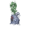
 分子量: 201.349 Da / 分子数: 1 / 由来タイプ: 組換発現 / 式: C12H27NO / タイプ: SUBJECT OF INVESTIGATION
 分子量: 1996.235 Da / 分子数: 1 / 由来タイプ: 合成 / 式: C93H164N2O39P2 / コメント: 毒素*YM
 試料調製
試料調製 電子顕微鏡撮影
 FIELD EMISSION GUN / 加速電圧: 300 kV / 照射モード: FLOOD BEAM
 解析
解析
