 Movie
Movie Controller

[English] 日本語
 Yorodumi
- PDB-8b14: T5 Receptor Binding Protein pb5 in complex with its E. coli recep... -
+ Open data

- Basic information
| Entry | Database: PDB / ID: 8b14 | |||||||||
|---|---|---|---|---|---|---|---|---|---|---|
| Title | T5 Receptor Binding Protein pb5 in complex with its E. coli receptor FhuA | |||||||||
 Components Components |
| |||||||||
 Keywords Keywords | VIRAL PROTEIN / bacteriophage / receptor / complex / RPB | |||||||||
| Function / homology |  Function and homology information Function and homology informationsiderophore transmembrane transport / siderophore uptake transmembrane transporter activity / virus tail / virion binding / toxic substance binding / transmembrane transporter complex / cell outer membrane / signaling receptor activity / intracellular iron ion homeostasis / entry receptor-mediated virion attachment to host cell ...siderophore transmembrane transport / siderophore uptake transmembrane transporter activity / virus tail / virion binding / toxic substance binding / transmembrane transporter complex / cell outer membrane / signaling receptor activity / intracellular iron ion homeostasis / entry receptor-mediated virion attachment to host cell / receptor-mediated virion attachment to host cell / iron ion binding / symbiont entry into host cell / protein domain specific binding / membrane Similarity search - Function | |||||||||
| Biological species |   Escherichia phage T5 (virus) Escherichia phage T5 (virus) | |||||||||
| Method | ELECTRON MICROSCOPY / single particle reconstruction / cryo EM / Resolution: 2.6 Å | |||||||||
 Authors Authors | Degroux, S. / Effantin, G. / Linares, R. / Schoehn, G. / Breyton, C. | |||||||||
| Funding support |  France, 2items France, 2items
| |||||||||
 Citation Citation | Journal: J Virol / Year: 2023 Title: Deciphering Bacteriophage T5 Host Recognition Mechanism and Infection Trigger. Authors: Séraphine Degroux / Grégory Effantin / Romain Linares / Guy Schoehn / Cécile Breyton / Abstract: Bacteriophages, viruses infecting bacteria, recognize their host with high specificity, binding to either saccharide motifs or proteins of the cell wall of their host. In the majority of ...Bacteriophages, viruses infecting bacteria, recognize their host with high specificity, binding to either saccharide motifs or proteins of the cell wall of their host. In the majority of bacteriophages, this host recognition is performed by receptor binding proteins (RBPs) located at the extremity of a tail. Interaction between the RBPs and the host is the trigger for bacteriophage infection, but the molecular details of the mechanisms are unknown for most bacteriophages. Here, we present the electron cryomicroscopy (cryo-EM) structure of bacteriophage T5 RBP in complex with its Escherichia coli receptor, the iron ferrichrome transporter FhuA. Monomeric RBP is located at the extremity of T5's long flexible tail, and its irreversible binding to FhuA commits T5 to infection. Analysis of the structure of RBP within the complex, comparison with its AlphaFold2-predicted structure, and its fit into a previously determined map of the T5 tail tip in complex with FhuA allow us to propose a mechanism of transmission of the RBP receptor binding to the straight fiber, initiating the cascade of events that commits T5 to DNA ejection. Tailed bacteriophages specifically recognize their bacterial host by interaction of their receptor binding protein(s) (RBPs) with saccharides and/or proteins located at the surface of their prey. This crucial interaction commits the virus to infection, but the molecular details of this mechanism are unknown for the majority of bacteriophages. We determined the structure of bacteriophage T5 RBP in complex with its E. coli receptor, FhuA, by cryo-EM. This first structure of an RBP bound to its protein receptor allowed us to propose a mechanism of transmission of host recognition to the rest of the phage, ultimately opening the capsid and perforating the cell wall and, thus, allowing safe channeling of the DNA into the host cytoplasm. | |||||||||
| History |
|
- Structure visualization
| Structure viewer | Molecule: MolmilJmol/JSmol |
|---|
- Downloads & links
-Download
| PDBx/mmCIF format | 8b14.cif.gz | 439.5 KB | Display | PDBx/mmCIF format |
|---|---|---|---|---|
| PDB format | pdb8b14.ent.gz | 363.7 KB | Display | PDB format |
| PDBx/mmJSON format | 8b14.json.gz | Tree view | PDBx/mmJSON format | |
| Others |  Other downloads Other downloads |
-Validation report
| Summary document | 8b14_validation.pdf.gz | 1.1 MB | Display | wwPDB validaton report |
|---|---|---|---|---|
| Full document | 8b14_full_validation.pdf.gz | 1.2 MB | Display | |
| Data in XML | 8b14_validation.xml.gz | 55.3 KB | Display | |
| Data in CIF | 8b14_validation.cif.gz | 82.2 KB | Display | |
| Arichive directory | https://data.pdbj.org/pub/pdb/validation_reports/b1/8b14ftp://data.pdbj.org/pub/pdb/validation_reports/b1/8b14 | HTTPS FTP |
-Related structure data
| Related structure data |  15802MC M: map data used to model this data C: citing same article ( |
|---|---|
| Similar structure data |
-Links
 PDBj
PDBj- Assembly
| Deposited unit | 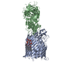
|
|---|---|
| 1 |
|
-Components
| #1: Protein | Mass: 79876.945 Da / Num. of mol.: 1 Mutation: Presence of an Histag after P405, added residues : SHHHHHHGS Source method: isolated from a genetically manipulated source Source: (gene. exp.) |
|---|---|
| #2: Protein | Mass: 68782.562 Da / Num. of mol.: 1 / Mutation: Presence of an Histag in C-ter position Source method: isolated from a genetically manipulated source Source: (gene. exp.) Escherichia phage T5 (virus) / Production host: |
| #3: Chemical | ChemComp-DDQ /   Mass: 201.349 Da / Num. of mol.: 1 Mass: 201.349 Da / Num. of mol.: 1Source method: isolated from a genetically manipulated source Formula: C12H27NO / Feature type: SUBJECT OF INVESTIGATION |
| #4: Chemical | ChemComp-LU9 / [(  Mass: 1996.235 Da / Num. of mol.: 1 / Source method: obtained synthetically / Formula: C93H164N2O39P2 / Comment: toxin*YM Mass: 1996.235 Da / Num. of mol.: 1 / Source method: obtained synthetically / Formula: C93H164N2O39P2 / Comment: toxin*YM |
| Has ligand of interest | Y |
-Experimental details
-Experiment
| Experiment | Method: ELECTRON MICROSCOPY |
|---|---|
| EM experiment | Aggregation state: PARTICLE / 3D reconstruction method: single particle reconstruction |
- Sample preparation
| Component |
| ||||||||||||||||||||||||||||
|---|---|---|---|---|---|---|---|---|---|---|---|---|---|---|---|---|---|---|---|---|---|---|---|---|---|---|---|---|---|
| Molecular weight | Value: 0.15 MDa / Experimental value: NO | ||||||||||||||||||||||||||||
| Source (natural) |
| ||||||||||||||||||||||||||||
| Source (recombinant) |
| ||||||||||||||||||||||||||||
| Buffer solution | pH: 8.5 | ||||||||||||||||||||||||||||
| Buffer component |
| ||||||||||||||||||||||||||||
| Specimen | Conc.: 1.1111111111111 mg/ml / Embedding applied: NO / Shadowing applied: NO / Staining applied: NO / Vitrification applied: YES Details: The FhuA-pb5 complex was formed by adding equimolar amounts of the two proteins, which results in 100% complex formation. FhuA-RBPpb5 complex is stabilized with 1.6% C10DAO at a protein ...Details: The FhuA-pb5 complex was formed by adding equimolar amounts of the two proteins, which results in 100% complex formation. FhuA-RBPpb5 complex is stabilized with 1.6% C10DAO at a protein concentration of 4.3 mg/ml | ||||||||||||||||||||||||||||
| Specimen support | Details: 25 mA / Grid material: COPPER/RHODIUM / Grid mesh size: 400 divisions/in. / Grid type: Quantifoil R2/1 | ||||||||||||||||||||||||||||
| Vitrification | Instrument: FEI VITROBOT MARK IV / Cryogen name: ETHANE / Humidity: 100 % / Chamber temperature: 293.15 K Details: 3.5 microliters of the FhuA-pb5 complex were deposited on a freshly glow discharged (25 mA, 30 sec) Cu/Rh 400 mesh Quantifoil R 2/1 EM grids and flash-frozen in nitrogen-cooled liquid ethane ...Details: 3.5 microliters of the FhuA-pb5 complex were deposited on a freshly glow discharged (25 mA, 30 sec) Cu/Rh 400 mesh Quantifoil R 2/1 EM grids and flash-frozen in nitrogen-cooled liquid ethane using a ThermoFisher Mark IV Vitrobot device (100% humidity, 293.15K, 2s blotting time, blot force 1). |
- Electron microscopy imaging
| Experimental equipment |  Model: Titan Krios / Image courtesy: FEI Company | ||||||||||||
|---|---|---|---|---|---|---|---|---|---|---|---|---|---|
| Microscopy | Model: FEI TITAN KRIOS / Details: calibrated pixel size = 1.052 | ||||||||||||
| Electron gun | Electron source:  FIELD EMISSION GUN / Accelerating voltage: 300 kV / Illumination mode: FLOOD BEAM FIELD EMISSION GUN / Accelerating voltage: 300 kV / Illumination mode: FLOOD BEAM | ||||||||||||
| Electron lens | Mode: BRIGHT FIELD / Nominal magnification: 130000 X / Nominal defocus max: 2600 nm / Nominal defocus min: 1200 nm / Cs: 2.7 mm / Alignment procedure: COMA FREE | ||||||||||||
| Specimen holder | Cryogen: NITROGEN / Specimen holder model: FEI TITAN KRIOS AUTOGRID HOLDER | ||||||||||||
| Image recording | Imaging-ID: 1 / Electron dose: 60 e/Å2 / Detector mode: COUNTING / Film or detector model: GATAN K2 SUMMIT (4k x 4k) / Num. of grids imaged: 1
| ||||||||||||
| EM imaging optics | Energyfilter name: GIF Quantum LS / Energyfilter slit width: 20 eV | ||||||||||||
| Image scans |
|
- Processing
| Software | Name: PHENIX / Version: 1.20.1_4487: / Classification: refinement | ||||||||||||||||||||||||||||||||
|---|---|---|---|---|---|---|---|---|---|---|---|---|---|---|---|---|---|---|---|---|---|---|---|---|---|---|---|---|---|---|---|---|---|
| EM software |
| ||||||||||||||||||||||||||||||||
| CTF correction | Type: PHASE FLIPPING AND AMPLITUDE CORRECTION | ||||||||||||||||||||||||||||||||
| Particle selection | Num. of particles selected: 2191586 | ||||||||||||||||||||||||||||||||
| Symmetry | Point symmetry: C1 (asymmetric) | ||||||||||||||||||||||||||||||||
| 3D reconstruction | Resolution: 2.6 Å / Resolution method: FSC 0.143 CUT-OFF / Num. of particles: 109350 / Algorithm: BACK PROJECTION / Symmetry type: POINT | ||||||||||||||||||||||||||||||||
| Atomic model building | Protocol: AB INITIO MODEL / Space: REAL Details: The pb5 protein model was built de novo in the cryo-electron microscopy map. FhuA was adapted from the FhuA structure solved by X-ray crystallography (PDB 2GRX). The two structures were ...Details: The pb5 protein model was built de novo in the cryo-electron microscopy map. FhuA was adapted from the FhuA structure solved by X-ray crystallography (PDB 2GRX). The two structures were first refined separately using Coot (version 0.9.2) and Phenix (version 1.18.2-3874) softwares, then together. Structure validation was done using MolProbity. | ||||||||||||||||||||||||||||||||
| Atomic model building | PDB-ID: 2GRX Accession code: 2GRX / Source name: PDB / Type: experimental model | ||||||||||||||||||||||||||||||||
| Refine LS restraints |
|

