Movie
Movie Controller


+ Open data

- Basic information
| Entry | Database: PDB / ID: 8avw | |||||||||
|---|---|---|---|---|---|---|---|---|---|---|
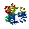
| Title | Cryo-EM structure of DrBphP in Pr state | |||||||||
 Components Components | Bacteriophytochrome,Response regulator | |||||||||
 Keywords Keywords | TRANSFERASE / KINASE / PHOTOSENSOR / PHYTOCHROME | |||||||||
| Function / homology |  Function and homology information Function and homology informationosmosensory signaling via phosphorelay pathway / detection of visible light / phosphorelay response regulator activity / protein kinase activator activity / histidine kinase / phosphorelay sensor kinase activity / photoreceptor activity / phosphorelay signal transduction system / regulation of DNA-templated transcription / ATP binding ...osmosensory signaling via phosphorelay pathway / detection of visible light / phosphorelay response regulator activity / protein kinase activator activity / histidine kinase / phosphorelay sensor kinase activity / photoreceptor activity / phosphorelay signal transduction system / regulation of DNA-templated transcription / ATP binding / identical protein binding / metal ion binding Similarity search - Function | |||||||||
| Biological species |  Deinococcus radiodurans R1 (radioresistant) Deinococcus radiodurans R1 (radioresistant) | |||||||||
| Method | ELECTRON MICROSCOPY / single particle reconstruction / cryo EM / Resolution: 3.62 Å | |||||||||
 Authors Authors | Wahlgren, W.Y. / Takala, H. / Westenhoff, S. | |||||||||
| Funding support | European Union,  Finland, 2items Finland, 2items
| |||||||||
 Citation Citation | Journal: Nat Commun / Year: 2022 Title: Structural mechanism of signal transduction in a phytochrome histidine kinase. Authors: Weixiao Yuan Wahlgren / Elin Claesson / Iida Tuure / Sergio Trillo-Muyo / Szabolcs Bódizs / Janne A Ihalainen / Heikki Takala / Sebastian Westenhoff /  Abstract: Phytochrome proteins detect red/far-red light to guide the growth, motion, development and reproduction in plants, fungi, and bacteria. Bacterial phytochromes commonly function as an entrance signal ...Phytochrome proteins detect red/far-red light to guide the growth, motion, development and reproduction in plants, fungi, and bacteria. Bacterial phytochromes commonly function as an entrance signal in two-component sensory systems. Despite the availability of three-dimensional structures of phytochromes and other two-component proteins, the conformational changes, which lead to activation of the protein, are not understood. We reveal cryo electron microscopy structures of the complete phytochrome from Deinoccocus radiodurans in its resting and photoactivated states at 3.6 Å and 3.5 Å resolution, respectively. Upon photoactivation, the photosensory core module hardly changes its tertiary domain arrangement, but the connector helices between the photosensory and the histidine kinase modules open up like a zipper, causing asymmetry and disorder in the effector domains. The structures provide a framework for atom-scale understanding of signaling in phytochromes, visualize allosteric communication over several nanometers, and suggest that disorder in the dimeric arrangement of the effector domains is important for phosphatase activity in a two-component system. The results have implications for the development of optogenetic applications. | |||||||||
| History |
|
- Structure visualization
| Structure viewer | Molecule: MolmilJmol/JSmol |
|---|
- Downloads & links
-Download
| PDBx/mmCIF format | 8avw.cif.gz | 246.2 KB | Display | PDBx/mmCIF format |
|---|---|---|---|---|
| PDB format | pdb8avw.ent.gz | 188.4 KB | Display | PDB format |
| PDBx/mmJSON format | 8avw.json.gz | Tree view | PDBx/mmJSON format | |
| Others |  Other downloads Other downloads |
-Validation report
| Summary document | 8avw_validation.pdf.gz | 1.5 MB | Display | wwPDB validaton report |
|---|---|---|---|---|
| Full document | 8avw_full_validation.pdf.gz | 1.6 MB | Display | |
| Data in XML | 8avw_validation.xml.gz | 52.2 KB | Display | |
| Data in CIF | 8avw_validation.cif.gz | 76.4 KB | Display | |
| Arichive directory | https://data.pdbj.org/pub/pdb/validation_reports/av/8avwftp://data.pdbj.org/pub/pdb/validation_reports/av/8avw | HTTPS FTP |
-Related structure data
| Related structure data |  15685MC 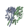 8avvC  8avxC M: map data used to model this data C: citing same article ( |
|---|---|
| Similar structure data |


-Links
 PDBj
PDBj


- Assembly
| Deposited unit | 
|
|---|---|
| 1 |
|
-Components
| #1: Protein | Mass: 101579.164 Da / Num. of mol.: 2 Source method: isolated from a genetically manipulated source Details: Sample is a fusion of Deinococcus radiodurans DrBphP and its response regulator DrRR.,Sample is a fusion of Deinococcus radiodurans DrBphP and its response regulator DrRR. Source: (gene. exp.) Deinococcus radiodurans R1 (radioresistant)Strain: ATCC 13939 / DSM 20539 / JCM 16871 / LMG 4051 / NBRC 15346 / NCIMB 9279 / R1 / VKM B-1422 Gene: bphP, DR_A0050, DR_A0049 / Plasmid: PET21B / Production host: References: UniProt: Q9RZA4, UniProt: Q9RZA5, histidine kinase #2: Chemical |   Mass: 585.670 Da / Num. of mol.: 2 / Source method: obtained synthetically / Formula: C33H37N4O6 / Feature type: SUBJECT OF INVESTIGATION Mass: 585.670 Da / Num. of mol.: 2 / Source method: obtained synthetically / Formula: C33H37N4O6 / Feature type: SUBJECT OF INVESTIGATIONHas ligand of interest | Y | |
|---|
-Experimental details
-Experiment
| Experiment | Method: ELECTRON MICROSCOPY |
|---|---|
| EM experiment | Aggregation state: PARTICLE / 3D reconstruction method: single particle reconstruction |
- Sample preparation
| Component | Name: Full-length bacteriophytochrome fused to its response regulator from Deinococcus radiodurans. Type: COMPLEX / Entity ID: #1 / Source: RECOMBINANT | ||||||||||||
|---|---|---|---|---|---|---|---|---|---|---|---|---|---|
| Molecular weight | Value: 0.101 MDa / Experimental value: NO | ||||||||||||
| Source (natural) |
| ||||||||||||
| Source (recombinant) |
| ||||||||||||
| Buffer solution | pH: 8 | ||||||||||||
| Specimen | Embedding applied: NO / Shadowing applied: NO / Staining applied: NO / Vitrification applied: YES | ||||||||||||
| Vitrification | Cryogen name: ETHANE |
- Electron microscopy imaging
| Experimental equipment |  Model: Titan Krios / Image courtesy: FEI Company |
|---|---|
| Microscopy | Model: FEI TITAN KRIOS |
| Electron gun | Electron source:  FIELD EMISSION GUN / Accelerating voltage: 300 kV / Illumination mode: FLOOD BEAM FIELD EMISSION GUN / Accelerating voltage: 300 kV / Illumination mode: FLOOD BEAM |
| Electron lens | Mode: BRIGHT FIELD / Nominal defocus max: 2600 nm / Nominal defocus min: 600 nm |
| Image recording | Electron dose: 48.3 e/Å2 / Film or detector model: GATAN K3 BIOQUANTUM (6k x 4k) |
- Processing
| EM software |
| ||||||||||||||||||||
|---|---|---|---|---|---|---|---|---|---|---|---|---|---|---|---|---|---|---|---|---|---|
| CTF correction | Type: NONE | ||||||||||||||||||||
| Symmetry | Point symmetry: C1 (asymmetric) | ||||||||||||||||||||
| 3D reconstruction | Resolution: 3.62 Å / Resolution method: FSC 0.143 CUT-OFF / Num. of particles: 117297 / Symmetry type: POINT | ||||||||||||||||||||
| Atomic model building | Protocol: RIGID BODY FIT / Space: REAL | ||||||||||||||||||||
| Atomic model building | PDB-ID: 4Q0J Pdb chain-ID: A / Accession code: 4Q0J / Source name: PDB / Type: experimental model |