 Movie
Movie Controller

[English] 日本語
 Yorodumi
- PDB-8ach: Nudaurelia capensis omega virus maturation intermediate captured ... -
+ Open data

- Basic information
| Entry | Database: PDB / ID: 8ach | |||||||||||||||||||||||||||||||||||||||||||||
|---|---|---|---|---|---|---|---|---|---|---|---|---|---|---|---|---|---|---|---|---|---|---|---|---|---|---|---|---|---|---|---|---|---|---|---|---|---|---|---|---|---|---|---|---|---|---|
| Title | Nudaurelia capensis omega virus maturation intermediate captured at pH5.6 (insect cell expressed VLPs): large class from symmetry expansion | |||||||||||||||||||||||||||||||||||||||||||||
 Components Components | p70 | |||||||||||||||||||||||||||||||||||||||||||||
 Keywords Keywords | VIRUS LIKE PARTICLE / ICOSAHEDRAL VIRUS / AUTO-CATALYTIC CLEAVAGE / VIRUS MATURATION / VIRUS-LIKE PARTICLE | |||||||||||||||||||||||||||||||||||||||||||||
| Function / homology | Peptidase N2 / Peptidase family A21 / virion component / Viral coat protein subunit / p70 Function and homology information Function and homology information | |||||||||||||||||||||||||||||||||||||||||||||
| Biological species |  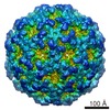 Nudaurelia capensis omega virus Nudaurelia capensis omega virus | |||||||||||||||||||||||||||||||||||||||||||||
| Method | ELECTRON MICROSCOPY / single particle reconstruction / cryo EM / Resolution: 3.91 Å | |||||||||||||||||||||||||||||||||||||||||||||
 Authors Authors | Castells-Graells, R. / Hesketh, E.L. / Johnson, J.E. / Ranson, N.A. / Lawson, D.M. / Lomonossoff, G.P. | |||||||||||||||||||||||||||||||||||||||||||||
| Funding support |  United Kingdom, 3items United Kingdom, 3items
| |||||||||||||||||||||||||||||||||||||||||||||
 Citation Citation | Journal: Proc Natl Acad Sci U S A / Year: 2026 Title: Unraveling the maturation pathway of a eukaryotic virus through cryo-EM. Authors: Roger Castells-Graells / Emma L Hesketh / Tsutomu Matsui / John E Johnson / Neil A Ranson / David M Lawson / George P Lomonossoff /  Abstract: Virus maturation is a fundamental biological process involving large-scale structural reorganizations that drive functional activation and lead to infectivity. Understanding the steps from the ...Virus maturation is a fundamental biological process involving large-scale structural reorganizations that drive functional activation and lead to infectivity. Understanding the steps from the initial procapsid assembly to mature virions is essential, both for comprehending viral life cycles and for developing antiviral therapies. However, capturing these steps has been challenging due to the transient and elusive nature of intermediate states. The nonenveloped, T = 4, ssRNA-containing, omega virus (NωV) is a highly accessible model system that exemplifies the maturation process of a eukaryotic virus. During maturation, the particle shrinks in outer diameter from 482 Å (pH 7.6) to 428 Å (pH 5.0). It is possible to mimic the maturation process in vitro by lowering the pH of a population of procapsids produced in heterologous systems. Indeed, by controlling the pH in vitro, it is possible to produce homogenous populations of intermediate NωV virus-like particles (VLPs) that occur too fleetingly to be observed in vivo. Here, we report structural models, based on cryoelectron microscopy (cryo-EM), of five intermediates in the NωV maturation process. The structures of the intermediate particles reveal unique, quaternary position-dependent trajectories and refolding of subunit N and C-terminal regions, including the formation of the autocatalytic cleavage site at N570. The detailed structures reported here, coupled with previously determined structures of the procapsids and mature particles, allow the maturation pathway to be described in detail for a eukaryotic virus. | |||||||||||||||||||||||||||||||||||||||||||||
| History |
|
- Structure visualization
| Structure viewer | Molecule: MolmilJmol/JSmol |
|---|
- Downloads & links
-Download
| PDBx/mmCIF format | 8ach.cif.gz | 368.1 KB | Display | PDBx/mmCIF format |
|---|---|---|---|---|
| PDB format | pdb8ach.ent.gz | 302.7 KB | Display | PDB format |
| PDBx/mmJSON format | 8ach.json.gz | Tree view | PDBx/mmJSON format | |
| Others |  Other downloads Other downloads |
-Validation report
| Arichive directory | https://data.pdbj.org/pub/pdb/validation_reports/ac/8achftp://data.pdbj.org/pub/pdb/validation_reports/ac/8ach | HTTPS FTP |
|---|
-Related structure data
| Related structure data |  15348MC  8a3cC  8a41C  8a6jC  8aayC  8ac6C M: map data used to model this data C: citing same article ( |
|---|---|
| Similar structure data |


-Links
 PDBj
PDBj
- Assembly
| Deposited unit | 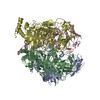
|
|---|---|
| 1 | x 60
|
-Components
| #1: Protein | Mass: 69891.953 Da / Num. of mol.: 4 Source method: isolated from a genetically manipulated source Source: (gene. exp.) Nudaurelia capensis omega virus / Cell (production host): Sf21 cell line / Production host:   Spodoptera frugiperda (fall armyworm) / References: UniProt: Q4TVS9 Spodoptera frugiperda (fall armyworm) / References: UniProt: Q4TVS9Has protein modification | N | |
|---|
-Experimental details
-Experiment
| Experiment | Method: ELECTRON MICROSCOPY |
|---|---|
| EM experiment | Aggregation state: PARTICLE / 3D reconstruction method: single particle reconstruction |
- Sample preparation
| Component | Name: Nudaurelia capensis omega virus / Type: VIRUS / Details: expressed in Spodoptera frugiperda Sf21 cells / Entity ID: all / Source: RECOMBINANT |
|---|---|
| Molecular weight | Value: 16.76 MDa / Experimental value: NO |
| Source (natural) | Organism: Nudaurelia capensis omega virus |
| Source (recombinant) | Organism: Spodoptera frugiperda (fall armyworm) / Cell: Sf21 |
| Details of virus | Empty: NO / Enveloped: NO / Isolate: OTHER / Type: VIRUS-LIKE PARTICLE |
| Natural host | Organism: Gonimbrasia cytherea |
| Virus shell | Name: coat / Diameter: 430 nm / Triangulation number (T number): 4 |
| Buffer solution | pH: 5.6 / Details: NULL |
| Specimen | Conc.: 0.3 mg/ml / Embedding applied: NO / Shadowing applied: NO / Staining applied: NO / Vitrification applied: YES / Details: NULL |
| Specimen support | Grid material: COPPER / Grid mesh size: 400 divisions/in. |
| Vitrification | Instrument: FEI VITROBOT MARK IV / Cryogen name: ETHANE |
- Electron microscopy imaging
| Experimental equipment |  Model: Titan Krios / Image courtesy: FEI Company |
|---|---|
| Microscopy | Model: FEI TITAN KRIOS |
| Electron gun | Electron source:  FIELD EMISSION GUN / Accelerating voltage: 300 kV / Illumination mode: FLOOD BEAM FIELD EMISSION GUN / Accelerating voltage: 300 kV / Illumination mode: FLOOD BEAM |
| Electron lens | Mode: BRIGHT FIELD / Nominal magnification: 75000 X / Nominal defocus max: 2500 nm / Nominal defocus min: 800 nm / Cs: 2.7 mm / C2 aperture diameter: 70 µm |
| Specimen holder | Cryogen: NITROGEN |
| Image recording | Average exposure time: 1 sec. / Electron dose: 49.1 e/Å2 / Detector mode: INTEGRATING / Film or detector model: FEI FALCON III (4k x 4k) / Num. of grids imaged: 2 / Num. of real images: 16478 Details: Reconstructed from 2 datasets collected at doses of 48.7 and 50.4 electrons per Angstrom squared, respectively. The average of these two values is given. |
- Processing
| EM software |
| ||||||||||||||||||||||||||||||||||||||||||||
|---|---|---|---|---|---|---|---|---|---|---|---|---|---|---|---|---|---|---|---|---|---|---|---|---|---|---|---|---|---|---|---|---|---|---|---|---|---|---|---|---|---|---|---|---|---|
| Image processing | Details: NULL | ||||||||||||||||||||||||||||||||||||||||||||
| CTF correction | Type: PHASE FLIPPING AND AMPLITUDE CORRECTION | ||||||||||||||||||||||||||||||||||||||||||||
| Symmetry | Point symmetry: C1 (asymmetric) | ||||||||||||||||||||||||||||||||||||||||||||
| 3D reconstruction | Resolution: 3.91 Å / Resolution method: FSC 0.143 CUT-OFF / Num. of particles: 96960 Details: Particles from an I1 icosahedral consensus map (EMD-15266) were symmetry expanded to C1 and further 3D classified without alignment using a soft mask covering the icosahedral asymmetric unit. ...Details: Particles from an I1 icosahedral consensus map (EMD-15266) were symmetry expanded to C1 and further 3D classified without alignment using a soft mask covering the icosahedral asymmetric unit. The particles for this class were randomly split into half sets for the subsequent reconstruction and postprocessing steps. Note that the symmetry expansion step generates a 60-fold larger particle stack. Symmetry type: POINT | ||||||||||||||||||||||||||||||||||||||||||||
| Atomic model building | B value: 161 / Protocol: OTHER / Space: REAL / Target criteria: Correlation coefficient | ||||||||||||||||||||||||||||||||||||||||||||
| Atomic model building | PDB-ID: 8AC6 Accession code: 8AC6 / Source name: PDB / Type: experimental model |
