 Movie
Movie Controller


+ Open data

- Basic information
| Entry | Database: PDB / ID: 7zqe | |||||||||||||||
|---|---|---|---|---|---|---|---|---|---|---|---|---|---|---|---|---|
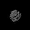
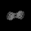
| Title | Plastocyanin bound to PSI of Chlamydomonas reinhardtii | |||||||||||||||
 Components Components | Plastocyanin, chloroplastic | |||||||||||||||
 Keywords Keywords | PHOTOSYNTHESIS / green algae / dimeric PSI | |||||||||||||||
| Function / homology |  Function and homology information Function and homology informationchloroplast thylakoid membrane / electron transfer activity / copper ion binding Similarity search - Function | |||||||||||||||
| Biological species |   Chlamydomonas reinhardtii (plant) Chlamydomonas reinhardtii (plant) | |||||||||||||||
| Method | ELECTRON MICROSCOPY / single particle reconstruction / cryo EM / Resolution: 2.55 Å | |||||||||||||||
 Authors Authors | Naschberger, A. / Amunts, A. | |||||||||||||||
| Funding support |  Sweden, Sweden,  Germany, 4items Germany, 4items
| |||||||||||||||
 Citation Citation | Journal: Nat Plants / Year: 2022 Title: Algal photosystem I dimer and high-resolution model of PSI-plastocyanin complex. Authors: Andreas Naschberger / Laura Mosebach / Victor Tobiasson / Sebastian Kuhlgert / Martin Scholz / Annemarie Perez-Boerema / Thi Thu Hoai Ho / André Vidal-Meireles / Yuichiro Takahashi / ...Authors: Andreas Naschberger / Laura Mosebach / Victor Tobiasson / Sebastian Kuhlgert / Martin Scholz / Annemarie Perez-Boerema / Thi Thu Hoai Ho / André Vidal-Meireles / Yuichiro Takahashi / Michael Hippler / Alexey Amunts /   Abstract: Photosystem I (PSI) enables photo-electron transfer and regulates photosynthesis in the bioenergetic membranes of cyanobacteria and chloroplasts. Being a multi-subunit complex, its macromolecular ...Photosystem I (PSI) enables photo-electron transfer and regulates photosynthesis in the bioenergetic membranes of cyanobacteria and chloroplasts. Being a multi-subunit complex, its macromolecular organization affects the dynamics of photosynthetic membranes. Here we reveal a chloroplast PSI from the green alga Chlamydomonas reinhardtii that is organized as a homodimer, comprising 40 protein subunits with 118 transmembrane helices that provide scaffold for 568 pigments. Cryogenic electron microscopy identified that the absence of PsaH and Lhca2 gives rise to a head-to-head relative orientation of the PSI-light-harvesting complex I monomers in a way that is essentially different from the oligomer formation in cyanobacteria. The light-harvesting protein Lhca9 is the key element for mediating this dimerization. The interface between the monomers is lacking PsaH and thus partially overlaps with the surface area that would bind one of the light-harvesting complex II complexes in state transitions. We also define the most accurate available PSI-light-harvesting complex I model at 2.3 Å resolution, including a flexibly bound electron donor plastocyanin, and assign correct identities and orientations to all the pigments, as well as 621 water molecules that affect energy transfer pathways. | |||||||||||||||
| History |
|
- Structure visualization
| Structure viewer | Molecule: MolmilJmol/JSmol |
|---|
- Downloads & links
-Download
| PDBx/mmCIF format | 7zqe.cif.gz | 42.5 KB | Display | PDBx/mmCIF format |
|---|---|---|---|---|
| PDB format | pdb7zqe.ent.gz | 28 KB | Display | PDB format |
| PDBx/mmJSON format | 7zqe.json.gz | Tree view | PDBx/mmJSON format | |
| Others |  Other downloads Other downloads |
-Validation report
| Arichive directory | https://data.pdbj.org/pub/pdb/validation_reports/zq/7zqeftp://data.pdbj.org/pub/pdb/validation_reports/zq/7zqe | HTTPS FTP |
|---|
-Related structure data
| Related structure data |  14872MC 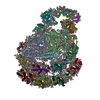 7zq9C 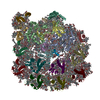 7zqcC 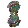 7zqdC M: map data used to model this data C: citing same article ( |
|---|---|
| Similar structure data |
-Links
 PDBj
PDBj- Assembly
| Deposited unit | 
|
|---|---|
| 1 |
|
-Components
| #1: Protein | Mass: 14889.784 Da / Num. of mol.: 1 / Source method: isolated from a natural source / Source: (natural) Chlamydomonas reinhardtii (plant) / References: UniProt: P18068 |
|---|---|
| #2: Chemical | ChemComp-CU /   Mass: 63.546 Da / Num. of mol.: 1 / Source method: obtained synthetically / Formula: Cu / Feature type: SUBJECT OF INVESTIGATION Mass: 63.546 Da / Num. of mol.: 1 / Source method: obtained synthetically / Formula: Cu / Feature type: SUBJECT OF INVESTIGATION |
| Has ligand of interest | Y |
-Experimental details
-Experiment
| Experiment | Method: ELECTRON MICROSCOPY |
|---|---|
| EM experiment | Aggregation state: PARTICLE / 3D reconstruction method: single particle reconstruction |
- Sample preparation
| Component | Name: Photosystem I dimer of Chlamydomonas reinhardtii / Type: ORGANELLE OR CELLULAR COMPONENT / Entity ID: #1 / Source: NATURAL |
|---|---|
| Molecular weight | Value: 0.7 MDa / Experimental value: YES |
| Source (natural) | Organism: Chlamydomonas reinhardtii (plant) |
| Buffer solution | pH: 7.5 |
| Specimen | Embedding applied: NO / Shadowing applied: NO / Staining applied: NO / Vitrification applied: YES |
| Vitrification | Cryogen name: ETHANE |
- Electron microscopy imaging
| Experimental equipment |  Model: Titan Krios / Image courtesy: FEI Company |
|---|---|
| Microscopy | Model: FEI TITAN KRIOS |
| Electron gun | Electron source:  FIELD EMISSION GUN / Accelerating voltage: 300 kV / Illumination mode: FLOOD BEAM FIELD EMISSION GUN / Accelerating voltage: 300 kV / Illumination mode: FLOOD BEAM |
| Electron lens | Mode: BRIGHT FIELD / Nominal defocus max: 5000 nm / Nominal defocus min: 1000 nm |
| Image recording | Electron dose: 45.8 e/Å2 / Film or detector model: GATAN K3 BIOQUANTUM (6k x 4k) |
- Processing
| CTF correction | Type: PHASE FLIPPING ONLY |
|---|---|
| 3D reconstruction | Resolution: 2.55 Å / Resolution method: FSC 0.143 CUT-OFF / Num. of particles: 66080 / Symmetry type: POINT |
