 Movie
Movie Controller

[English] 日本語
 Yorodumi
- PDB-7yrn: Cyro-EM structure of HCMV glycoprotein B in complex with 1B03 Fab -
+ Open data

- Basic information
| Entry | Database: PDB / ID: 7yrn | |||||||||||||||||||||||||||||||||
|---|---|---|---|---|---|---|---|---|---|---|---|---|---|---|---|---|---|---|---|---|---|---|---|---|---|---|---|---|---|---|---|---|---|---|
| Title | Cyro-EM structure of HCMV glycoprotein B in complex with 1B03 Fab | |||||||||||||||||||||||||||||||||
 Components Components |
| |||||||||||||||||||||||||||||||||
 Keywords Keywords | VIRAL PROTEIN/IMMUNE SYSTEM / VIRAL PROTEN / VIRAL PROTEIN-IMMUNE SYSTEM complex | |||||||||||||||||||||||||||||||||
| Function / homology |  Function and homology information Function and homology informationhost cell endosome / host cell Golgi apparatus / viral envelope / symbiont entry into host cell / virion attachment to host cell / host cell plasma membrane / membrane Similarity search - Function | |||||||||||||||||||||||||||||||||
| Biological species |   Human betaherpesvirus 5 Human betaherpesvirus 5 Homo sapiens (human) Homo sapiens (human) | |||||||||||||||||||||||||||||||||
| Method | ELECTRON MICROSCOPY / single particle reconstruction / cryo EM / Resolution: 2.99 Å | |||||||||||||||||||||||||||||||||
 Authors Authors | Wang, H. / Zhu, S. / Liao, H. | |||||||||||||||||||||||||||||||||
| Funding support |  China, 1items China, 1items
| |||||||||||||||||||||||||||||||||
 Citation Citation | Journal: To Be Published Title: Cyro-EM structure of HCMV glycoprotein B in complex with 1B03 Fab Authors: Wu, C. / Wang, H. / Zhu, S. / Liao, H. | |||||||||||||||||||||||||||||||||
| History |
|
- Structure visualization
| Structure viewer | Molecule: MolmilJmol/JSmol |
|---|
- Downloads & links
-Download
| PDBx/mmCIF format | 7yrn.cif.gz | 427.8 KB | Display | PDBx/mmCIF format |
|---|---|---|---|---|
| PDB format | pdb7yrn.ent.gz | Display | PDB format | |
| PDBx/mmJSON format | 7yrn.json.gz | Tree view | PDBx/mmJSON format | |
| Others |  Other downloads Other downloads |
-Validation report
| Summary document | 7yrn_validation.pdf.gz | 1.4 MB | Display | wwPDB validaton report |
|---|---|---|---|---|
| Full document | 7yrn_full_validation.pdf.gz | 1.4 MB | Display | |
| Data in XML | 7yrn_validation.xml.gz | 65.9 KB | Display | |
| Data in CIF | 7yrn_validation.cif.gz | 95.8 KB | Display | |
| Arichive directory | https://data.pdbj.org/pub/pdb/validation_reports/yr/7yrnftp://data.pdbj.org/pub/pdb/validation_reports/yr/7yrn | HTTPS FTP |
-Related structure data
| Related structure data | 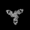 34063MC M: map data used to model this data C: citing same article ( |
|---|---|
| Similar structure data |
-Links
 PDBj
PDBj

- Assembly
| Deposited unit | 
|
|---|---|
| 1 |
|
-Components
| #1: Protein | Mass: 83545.547 Da / Num. of mol.: 3 Source method: isolated from a genetically manipulated source Source: (gene. exp.) Human betaherpesvirus 5 / Strain: Towne / Gene: UL55, gB / Cell line (production host): HEK293 / Production host: Homo sapiens (human) / References: UniProt: B9VXM4#2: Antibody | Mass: 23851.783 Da / Num. of mol.: 3 Source method: isolated from a genetically manipulated source Source: (gene. exp.) Homo sapiens (human) / Cell line (production host): HEK293 / Production host: Homo sapiens (human)#3: Antibody | Mass: 23508.117 Da / Num. of mol.: 3 Source method: isolated from a genetically manipulated source Source: (gene. exp.) Homo sapiens (human) / Cell line (production host): HEK293 / Production host: Homo sapiens (human)#4: Polysaccharide | Source method: isolated from a genetically manipulated source #5: Sugar | ChemComp-NAG /   Type: D-saccharide, beta linking / Mass: 221.208 Da / Num. of mol.: 6 / Source method: obtained synthetically / Formula: C8H15NO6 / Feature type: SUBJECT OF INVESTIGATION Type: D-saccharide, beta linking / Mass: 221.208 Da / Num. of mol.: 6 / Source method: obtained synthetically / Formula: C8H15NO6 / Feature type: SUBJECT OF INVESTIGATIONHas ligand of interest | Y | Has protein modification | Y | |
|---|
-Experimental details
-Experiment
| Experiment | Method: ELECTRON MICROSCOPY |
|---|---|
| EM experiment | Aggregation state: PARTICLE / 3D reconstruction method: single particle reconstruction |
- Sample preparation
| Component |
| ||||||||||||||||||||||||
|---|---|---|---|---|---|---|---|---|---|---|---|---|---|---|---|---|---|---|---|---|---|---|---|---|---|
| Source (natural) |
| ||||||||||||||||||||||||
| Source (recombinant) |
| ||||||||||||||||||||||||
| Buffer solution | pH: 8 | ||||||||||||||||||||||||
| Specimen | Embedding applied: NO / Shadowing applied: NO / Staining applied: NO / Vitrification applied: YES | ||||||||||||||||||||||||
| Vitrification | Cryogen name: ETHANE / Humidity: 100 % |
- Electron microscopy imaging
| Experimental equipment |  Model: Titan Krios / Image courtesy: FEI Company |
|---|---|
| Microscopy | Model: FEI TITAN KRIOS |
| Electron gun | Electron source:  FIELD EMISSION GUN / Accelerating voltage: 300 kV / Illumination mode: FLOOD BEAM FIELD EMISSION GUN / Accelerating voltage: 300 kV / Illumination mode: FLOOD BEAM |
| Electron lens | Mode: BRIGHT FIELD / Nominal defocus max: 2500 nm / Nominal defocus min: 1000 nm |
| Image recording | Electron dose: 60 e/Å2 / Film or detector model: GATAN K3 (6k x 4k) |
- Processing
| Software | Name: PHENIX / Version: 1.20_4459: / Classification: refinement | ||||||||||||||||||||||||
|---|---|---|---|---|---|---|---|---|---|---|---|---|---|---|---|---|---|---|---|---|---|---|---|---|---|
| EM software | Name: PHENIX / Category: model refinement | ||||||||||||||||||||||||
| CTF correction | Type: PHASE FLIPPING AND AMPLITUDE CORRECTION | ||||||||||||||||||||||||
| 3D reconstruction | Resolution: 2.99 Å / Resolution method: FSC 0.143 CUT-OFF / Num. of particles: 185463 / Symmetry type: POINT | ||||||||||||||||||||||||
| Refine LS restraints |
|
