 ムービー
ムービー コントローラー


+ データを開く

- 基本情報
基本情報
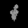
| 登録情報 | データベース: PDB / ID: 7y9s | |||||||||||||||||||||||||||||||||||||||||||||
|---|---|---|---|---|---|---|---|---|---|---|---|---|---|---|---|---|---|---|---|---|---|---|---|---|---|---|---|---|---|---|---|---|---|---|---|---|---|---|---|---|---|---|---|---|---|---|
| タイトル | Cryo-EM structure of apo SARS-CoV-2 Omicron spike protein (S-2P-GSAS) | |||||||||||||||||||||||||||||||||||||||||||||
 要素 要素 | Spike glycoprotein | |||||||||||||||||||||||||||||||||||||||||||||
 キーワード キーワード | VIRAL PROTEIN / SARS-CoV-2 / Omicron / spike protein | |||||||||||||||||||||||||||||||||||||||||||||
| 機能・相同性 |  機能・相同性情報 機能・相同性情報symbiont-mediated disruption of host tissue / Maturation of spike protein / Translation of Structural Proteins / Virion Assembly and Release / host cell surface / host extracellular region / symbiont-mediated-mediated suppression of host tetherin activity / Induction of Cell-Cell Fusion / structural constituent of virion / positive regulation of viral entry into host cell ...symbiont-mediated disruption of host tissue / Maturation of spike protein / Translation of Structural Proteins / Virion Assembly and Release / host cell surface / host extracellular region / symbiont-mediated-mediated suppression of host tetherin activity / Induction of Cell-Cell Fusion / structural constituent of virion / positive regulation of viral entry into host cell / membrane fusion / host cell endoplasmic reticulum-Golgi intermediate compartment membrane / Attachment and Entry / entry receptor-mediated virion attachment to host cell / receptor-mediated virion attachment to host cell / host cell surface receptor binding / symbiont-mediated suppression of host innate immune response / endocytosis involved in viral entry into host cell / receptor ligand activity / fusion of virus membrane with host plasma membrane / fusion of virus membrane with host endosome membrane / viral envelope / symbiont entry into host cell / virion attachment to host cell / host cell plasma membrane / SARS-CoV-2 activates/modulates innate and adaptive immune responses / virion membrane / membrane / identical protein binding / plasma membrane 類似検索 - 分子機能 | |||||||||||||||||||||||||||||||||||||||||||||
| 生物種 |   Severe acute respiratory syndrome coronavirus 2 (ウイルス) Severe acute respiratory syndrome coronavirus 2 (ウイルス) | |||||||||||||||||||||||||||||||||||||||||||||
| 手法 | 電子顕微鏡法 / 単粒子再構成法 / クライオ電子顕微鏡法 / 解像度: 3 Å | |||||||||||||||||||||||||||||||||||||||||||||
 データ登録者 データ登録者 | Zhao, Z.N. / Xie, Y.F. / Qi, J.X. / Gao, G.F. | |||||||||||||||||||||||||||||||||||||||||||||
| 資金援助 |  中国, 1件 中国, 1件
| |||||||||||||||||||||||||||||||||||||||||||||
 引用 引用 | ジャーナル: Nat Commun / 年: 2022 タイトル: Omicron SARS-CoV-2 mutations stabilize spike up-RBD conformation and lead to a non-RBM-binding monoclonal antibody escape. 著者: Zhennan Zhao / Jingya Zhou / Mingxiong Tian / Min Huang / Sheng Liu / Yufeng Xie / Pu Han / Chongzhi Bai / Pengcheng Han / Anqi Zheng / Lutang Fu / Yuanzhu Gao / Qi Peng / Ying Li / Yan Chai ...著者: Zhennan Zhao / Jingya Zhou / Mingxiong Tian / Min Huang / Sheng Liu / Yufeng Xie / Pu Han / Chongzhi Bai / Pengcheng Han / Anqi Zheng / Lutang Fu / Yuanzhu Gao / Qi Peng / Ying Li / Yan Chai / Zengyuan Zhang / Xin Zhao / Hao Song / Jianxun Qi / Qihui Wang / Peiyi Wang / George F Gao / 要旨: Omicron SARS-CoV-2 is rapidly spreading worldwide. To delineate the impact of emerging mutations on spike's properties, we performed systematic structural analyses on apo Omicron spike and its ...Omicron SARS-CoV-2 is rapidly spreading worldwide. To delineate the impact of emerging mutations on spike's properties, we performed systematic structural analyses on apo Omicron spike and its complexes with human ACE2 or S309 neutralizing antibody (NAb) by cryo-EM. The Omicron spike preferentially adopts the one-RBD-up conformation both before and after ACE2 binding, which is in sharp contrast to the orchestrated conformational changes to create more up-RBDs upon ACE2 binding as observed in the prototype and other four variants of concern (VOCs). Furthermore, we found that S371L, S373P and S375F substitutions enhance the stability of the one-RBD-up conformation to prevent exposing more up-RBDs triggered by ACE2 binding. The increased stability of the one-RBD-up conformation restricts the accessibility of S304 NAb, which targets a cryptic epitope in the closed conformation, thus facilitating the immune evasion by Omicron. These results expand our understanding of Omicron spike's conformation, receptor binding and antibody evasion mechanism. | |||||||||||||||||||||||||||||||||||||||||||||
| 履歴 |
|
- 構造の表示
構造の表示
| 構造ビューア | 分子: MolmilJmol/JSmol |
|---|
- ダウンロードとリンク
-ダウンロード
| PDBx/mmCIF形式 | 7y9s.cif.gz | 638.9 KB | 表示 | PDBx/mmCIF形式 |
|---|---|---|---|---|
| PDB形式 | pdb7y9s.ent.gz | 515.2 KB | 表示 | PDB形式 |
| PDBx/mmJSON形式 | 7y9s.json.gz | ツリー表示 | PDBx/mmJSON形式 | |
| その他 |  その他のダウンロード その他のダウンロード |
-検証レポート
| アーカイブディレクトリ | https://data.pdbj.org/pub/pdb/validation_reports/y9/7y9sftp://data.pdbj.org/pub/pdb/validation_reports/y9/7y9s | HTTPS FTP |
|---|
-関連構造データ


-リンク
 PDBj
PDBj


- 集合体
集合体
| 登録構造単位 | 
|
|---|---|
| 1 |
|
-要素
| #1: タンパク質 | 分子量: 138842.375 Da / 分子数: 3 / 変異: K986P, V987P / 由来タイプ: 組換発現 由来: (組換発現) Severe acute respiratory syndrome coronavirus 2 (ウイルス)遺伝子: S, 2 / 発現宿主:  Homo sapiens (ヒト) / 参照: UniProt: P0DTC2 Homo sapiens (ヒト) / 参照: UniProt: P0DTC2#2: 多糖 | 2-acetamido-2-deoxy-beta-D-glucopyranose-(1-4)-2-acetamido-2-deoxy-beta-D-glucopyranose #3: 糖 | ChemComp-NAG /   タイプ: D-saccharide, beta linking / 分子量: 221.208 Da / 分子数: 21 / 由来タイプ: 合成 / 式: C8H15NO6 / タイプ: SUBJECT OF INVESTIGATION タイプ: D-saccharide, beta linking / 分子量: 221.208 Da / 分子数: 21 / 由来タイプ: 合成 / 式: C8H15NO6 / タイプ: SUBJECT OF INVESTIGATION研究の焦点であるリガンドがあるか | Y | Has protein modification | Y | |
|---|
-実験情報
-実験
| 実験 | 手法: 電子顕微鏡法 |
|---|---|
| EM実験 | 試料の集合状態: PARTICLE / 3次元再構成法: 単粒子再構成法 |
- 試料調製
試料調製
| 構成要素 | 名称: Cryo-EM structure of SARS-CoV-2 Omicron spike protein タイプ: COMPLEX / Entity ID: #1 / 由来: RECOMBINANT |
|---|---|
| 由来(天然) | 生物種: Severe acute respiratory syndrome coronavirus 2 (ウイルス) |
| 由来(組換発現) | 生物種: Homo sapiens (ヒト) |
| 緩衝液 | pH: 8 |
| 試料 | 包埋: NO / シャドウイング: NO / 染色: NO / 凍結: YES |
| 急速凍結 | 凍結剤: ETHANE |
- 電子顕微鏡撮影
| 実験機器 |  モデル: Titan Krios / 画像提供: FEI Company |
|---|---|
| 顕微鏡 | モデル: FEI TITAN KRIOS |
| 電子銃 | 電子線源:  FIELD EMISSION GUN / 加速電圧: 300 kV / 照射モード: FLOOD BEAM FIELD EMISSION GUN / 加速電圧: 300 kV / 照射モード: FLOOD BEAM |
| 電子レンズ | モード: BRIGHT FIELD / 最大 デフォーカス(公称値): 5000 nm / 最小 デフォーカス(公称値): 1200 nm |
| 撮影 | 電子線照射量: 60 e/Å2 フィルム・検出器のモデル: GATAN K3 BIOQUANTUM (6k x 4k) |
- 解析
解析
| ソフトウェア | 名称: PHENIX / バージョン: 1.20.1_4487: / 分類: 精密化 | ||||||||||||||||||||||||
|---|---|---|---|---|---|---|---|---|---|---|---|---|---|---|---|---|---|---|---|---|---|---|---|---|---|
| EMソフトウェア | 名称: PHENIX / カテゴリ: モデル精密化 | ||||||||||||||||||||||||
| CTF補正 | タイプ: NONE | ||||||||||||||||||||||||
| 3次元再構成 | 解像度: 3 Å / 解像度の算出法: FSC 0.143 CUT-OFF / 粒子像の数: 98093 / 対称性のタイプ: POINT | ||||||||||||||||||||||||
| 拘束条件 |
|
