 Movie
Movie Controller


+ Open data

- Basic information
| Entry | Database: PDB / ID: 7y89 | |||||||||
|---|---|---|---|---|---|---|---|---|---|---|
| Title | Structure of the GPR17-Gi complex | |||||||||
 Components Components |
| |||||||||
 Keywords Keywords | MEMBRANE PROTEIN / classA GPCR / complex | |||||||||
| Function / homology |  Function and homology information Function and homology information: / Leukotriene receptors / P2Y receptors / chemokine receptor activity / receptor serine/threonine kinase binding / positive regulation of Rho protein signal transduction / oligodendrocyte differentiation / plasma membrane => GO:0005886 / Adenylate cyclase inhibitory pathway / positive regulation of protein localization to cell cortex ...: / Leukotriene receptors / P2Y receptors / chemokine receptor activity / receptor serine/threonine kinase binding / positive regulation of Rho protein signal transduction / oligodendrocyte differentiation / plasma membrane => GO:0005886 / Adenylate cyclase inhibitory pathway / positive regulation of protein localization to cell cortex / regulation of cAMP-mediated signaling / D2 dopamine receptor binding / G protein-coupled serotonin receptor binding / regulation of mitotic spindle organization / cellular response to forskolin / negative regulation of inflammatory response to antigenic stimulus / adenylate cyclase-inhibiting G protein-coupled receptor signaling pathway / Regulation of insulin secretion / G protein-coupled receptor binding / G protein-coupled receptor activity / G-protein beta/gamma-subunit complex binding / Olfactory Signaling Pathway / Activation of the phototransduction cascade / adenylate cyclase-modulating G protein-coupled receptor signaling pathway / G beta:gamma signalling through PLC beta / Presynaptic function of Kainate receptors / Thromboxane signalling through TP receptor / G-protein activation / G protein-coupled acetylcholine receptor signaling pathway / Activation of G protein gated Potassium channels / Inhibition of voltage gated Ca2+ channels via Gbeta/gamma subunits / Prostacyclin signalling through prostacyclin receptor / Glucagon signaling in metabolic regulation / G beta:gamma signalling through CDC42 / ADP signalling through P2Y purinoceptor 12 / G beta:gamma signalling through BTK / response to peptide hormone / Synthesis, secretion, and inactivation of Glucagon-like Peptide-1 (GLP-1) / Sensory perception of sweet, bitter, and umami (glutamate) taste / photoreceptor disc membrane / Adrenaline,noradrenaline inhibits insulin secretion / Glucagon-type ligand receptors / Vasopressin regulates renal water homeostasis via Aquaporins / G alpha (z) signalling events / cellular response to catecholamine stimulus / Glucagon-like Peptide-1 (GLP1) regulates insulin secretion / ADORA2B mediated anti-inflammatory cytokines production / sensory perception of taste / ADP signalling through P2Y purinoceptor 1 / adenylate cyclase-activating dopamine receptor signaling pathway / G beta:gamma signalling through PI3Kgamma / cellular response to prostaglandin E stimulus / Cooperation of PDCL (PhLP1) and TRiC/CCT in G-protein beta folding / GPER1 signaling / GDP binding / Inactivation, recovery and regulation of the phototransduction cascade / heterotrimeric G-protein complex / G alpha (12/13) signalling events / extracellular vesicle / signaling receptor complex adaptor activity / Thrombin signalling through proteinase activated receptors (PARs) / GTPase binding / retina development in camera-type eye / phospholipase C-activating G protein-coupled receptor signaling pathway / Ca2+ pathway / cell cortex / midbody / G alpha (i) signalling events / G alpha (s) signalling events / G alpha (q) signalling events / cell population proliferation / Ras protein signal transduction / Extra-nuclear estrogen signaling / cell cycle / G protein-coupled receptor signaling pathway / cell division / lysosomal membrane / GTPase activity / centrosome / synapse / protein-containing complex binding / nucleolus / GTP binding / magnesium ion binding / signal transduction / extracellular exosome / nucleoplasm / membrane / plasma membrane / cytoplasm / cytosol Similarity search - Function | |||||||||
| Biological species |  Homo sapiens (human) Homo sapiens (human) | |||||||||
| Method | ELECTRON MICROSCOPY / single particle reconstruction / cryo EM / Resolution: 3.02 Å | |||||||||
 Authors Authors | Ye, F. / Chen, G. | |||||||||
| Funding support | 1items
| |||||||||
 Citation Citation | Journal: MedComm (2020) / Year: 2022 Title: Cryo-EM structure of G-protein-coupled receptor GPR17 in complex with inhibitory G protein. Authors: Fang Ye / Thian-Sze Wong / Geng Chen / Zhiyi Zhang / Binghao Zhang / Shiyi Gan / Wei Gao / Jiancheng Li / Zhangsong Wu / Xin Pan / Yang Du /  Abstract: GPR17 is a class A orphan G protein-coupled receptor (GPCR) expressed in neurons and oligodendrocyte progenitors of the central nervous system (CNS). The signalling of GPR17 occurs through the ...GPR17 is a class A orphan G protein-coupled receptor (GPCR) expressed in neurons and oligodendrocyte progenitors of the central nervous system (CNS). The signalling of GPR17 occurs through the heterotrimeric Gi, but its activation mechanism is unclear. Here, we employed cryo-electron microscopy (cryo-EM) technology to elucidate the structure of activated GPR17-Gi complex. The 3.02 Å resolution structure, together with mutagenesis studies, revealed that the extracellular loop2 of GPR17 occupied the orthosteric binding pocket to promote its self-activation. The active GPR17 carried several typical microswitches like other class A GPCRs. Moreover, the Gi interacted with the key residues of transmembrane helix 3 (TM3), the amphipathic helix 8 (Helix8), and intracellular loops 3 (ICL3) in GPR17 to engage in the receptor core. In summary, our results highlight the activation mechanism of GPR17 from the structural basis. Elucidating the structural and activation mechanism of GPR17 may facilitate the pharmacological intervention for acute/chronic CNS injury. | |||||||||
| History |
|
- Structure visualization
| Structure viewer | Molecule: MolmilJmol/JSmol |
|---|
- Downloads & links
-Download
| PDBx/mmCIF format | 7y89.cif.gz | 203.1 KB | Display | PDBx/mmCIF format |
|---|---|---|---|---|
| PDB format | pdb7y89.ent.gz | 160.6 KB | Display | PDB format |
| PDBx/mmJSON format | 7y89.json.gz | Tree view | PDBx/mmJSON format | |
| Others |  Other downloads Other downloads |
-Validation report
| Summary document | 7y89_validation.pdf.gz | 949 KB | Display | wwPDB validaton report |
|---|---|---|---|---|
| Full document | 7y89_full_validation.pdf.gz | 965.7 KB | Display | |
| Data in XML | 7y89_validation.xml.gz | 41.6 KB | Display | |
| Data in CIF | 7y89_validation.cif.gz | 62.4 KB | Display | |
| Arichive directory | https://data.pdbj.org/pub/pdb/validation_reports/y8/7y89ftp://data.pdbj.org/pub/pdb/validation_reports/y8/7y89 | HTTPS FTP |
-Related structure data
| Related structure data | 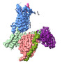 33682MC M: map data used to model this data C: citing same article ( |
|---|---|
| Similar structure data |
-Links
 PDBj
PDBj


- Assembly
| Deposited unit | 
|
|---|---|
| 1 |
|
-Components
| #1: Protein | Mass: 40153.672 Da / Num. of mol.: 1 / Source method: isolated from a natural source / Source: (natural) Homo sapiens (human) / References: UniProt: P63096 |
|---|---|
| #2: Protein | Mass: 37069.543 Da / Num. of mol.: 1 / Source method: isolated from a natural source / Source: (natural) Homo sapiens (human) / References: UniProt: P62873 |
| #3: Protein | Mass: 6218.162 Da / Num. of mol.: 1 / Source method: isolated from a natural source / Source: (natural) Homo sapiens (human) |
| #4: Antibody | Mass: 26424.385 Da / Num. of mol.: 1 / Source method: isolated from a natural source / Source: (natural) Homo sapiens (human) |
| #5: Protein | Mass: 33067.391 Da / Num. of mol.: 1 / Source method: isolated from a natural source / Source: (natural) |
-Experimental details
-Experiment
| Experiment | Method: ELECTRON MICROSCOPY |
|---|---|
| EM experiment | Aggregation state: CELL / 3D reconstruction method: single particle reconstruction |
- Sample preparation
| Component | Name: GPR17-Gi / Type: COMPLEX / Entity ID: all / Source: NATURAL |
|---|---|
| Source (natural) | Organism: Homo sapiens (human) |
| Buffer solution | pH: 7.4 |
| Specimen | Embedding applied: NO / Shadowing applied: NO / Staining applied: NO / Vitrification applied: YES |
| Vitrification | Cryogen name: ETHANE |
- Electron microscopy imaging
| Experimental equipment |  Model: Titan Krios / Image courtesy: FEI Company |
|---|---|
| Microscopy | Model: FEI TITAN KRIOS |
| Electron gun | Electron source:  FIELD EMISSION GUN / Accelerating voltage: 300 kV / Illumination mode: SPOT SCAN FIELD EMISSION GUN / Accelerating voltage: 300 kV / Illumination mode: SPOT SCAN |
| Electron lens | Mode: BRIGHT FIELD / Nominal defocus max: 1400 nm / Nominal defocus min: 1000 nm |
| Image recording | Electron dose: 57 e/Å2 / Film or detector model: GATAN K3 BIOQUANTUM (6k x 4k) |
- Processing
| Software | Name: PHENIX / Version: 1.18.2_3874: / Classification: refinement | ||||||||||||||||||||||||
|---|---|---|---|---|---|---|---|---|---|---|---|---|---|---|---|---|---|---|---|---|---|---|---|---|---|
| CTF correction | Type: PHASE FLIPPING AND AMPLITUDE CORRECTION | ||||||||||||||||||||||||
| 3D reconstruction | Resolution: 3.02 Å / Resolution method: FSC 0.143 CUT-OFF / Num. of particles: 314674 / Symmetry type: POINT | ||||||||||||||||||||||||
| Refine LS restraints |
|
