 Movie
Movie Controller

[English] 日本語
 Yorodumi
- PDB-7x9a: Cryo-EM structure of neuropeptide Y Y1 receptor in complex with N... -
+ Open data

- Basic information
| Entry | Database: PDB / ID: 7x9a | ||||||||||||
|---|---|---|---|---|---|---|---|---|---|---|---|---|---|
| Title | Cryo-EM structure of neuropeptide Y Y1 receptor in complex with NPY and Gi | ||||||||||||
 Components Components |
| ||||||||||||
 Keywords Keywords | SIGNALING PROTEIN | ||||||||||||
| Function / homology |  Function and homology information Function and homology informationpancreatic polypeptide receptor activity / regulation of nerve growth factor production / peptide YY receptor activity / short-day photoperiodism / negative regulation of acute inflammatory response to antigenic stimulus / neuropeptide Y receptor activity / monocyte activation / positive regulation of dopamine metabolic process / : / neuropeptide Y receptor binding ...pancreatic polypeptide receptor activity / regulation of nerve growth factor production / peptide YY receptor activity / short-day photoperiodism / negative regulation of acute inflammatory response to antigenic stimulus / neuropeptide Y receptor activity / monocyte activation / positive regulation of dopamine metabolic process / : / neuropeptide Y receptor binding / intestinal epithelial cell differentiation / positive regulation of appetite / positive regulation of eating behavior / synaptic signaling via neuropeptide / adult feeding behavior / neuropeptide receptor activity / regulation of presynaptic cytosolic calcium ion concentration / drinking behavior / neuropeptide hormone activity / positive regulation of cell-substrate adhesion / feeding behavior / neuropeptide binding / regulation of synaptic vesicle exocytosis / central nervous system neuron development / outflow tract morphogenesis / regulation of multicellular organism growth / developmental growth / FOXO-mediated transcription of oxidative stress, metabolic and neuronal genes / G protein-coupled receptor signaling pathway, coupled to cyclic nucleotide second messenger / neuronal dense core vesicle / negative regulation of blood pressure / adenylate cyclase inhibitor activity / positive regulation of protein localization to cell cortex / T cell migration / positive regulation of relaxation of smooth muscle / Adenylate cyclase inhibitory pathway / D2 dopamine receptor binding / sensory perception of pain / adenylate cyclase-inhibiting serotonin receptor signaling pathway / G protein-coupled serotonin receptor binding / cellular response to forskolin / Peptide ligand-binding receptors / regulation of mitotic spindle organization / chemokine-mediated signaling pathway / calcium channel regulator activity / Regulation of insulin secretion / neuropeptide signaling pathway / response to prostaglandin E / locomotory behavior / cerebral cortex development / positive regulation of cholesterol biosynthetic process / negative regulation of insulin secretion / G protein-coupled receptor binding / response to peptide hormone / regulation of blood pressure / GABA-ergic synapse / glucose metabolic process / G protein-coupled receptor activity / centriolar satellite / G-protein beta/gamma-subunit complex binding / adenylate cyclase-modulating G protein-coupled receptor signaling pathway / adenylate cyclase-inhibiting G protein-coupled receptor signaling pathway / neuron projection development / Olfactory Signaling Pathway / Activation of the phototransduction cascade / terminal bouton / G protein-coupled acetylcholine receptor signaling pathway / G beta:gamma signalling through PLC beta / Presynaptic function of Kainate receptors / Thromboxane signalling through TP receptor / Activation of G protein gated Potassium channels / Inhibition of voltage gated Ca2+ channels via Gbeta/gamma subunits / G-protein activation / Glucagon signaling in metabolic regulation / Prostacyclin signalling through prostacyclin receptor / G beta:gamma signalling through CDC42 / Synthesis, secretion, and inactivation of Glucagon-like Peptide-1 (GLP-1) / G beta:gamma signalling through BTK / photoreceptor disc membrane / ADP signalling through P2Y purinoceptor 12 / Glucagon-type ligand receptors / Sensory perception of sweet, bitter, and umami (glutamate) taste / GDP binding / Adrenaline,noradrenaline inhibits insulin secretion / Vasopressin regulates renal water homeostasis via Aquaporins / Glucagon-like Peptide-1 (GLP1) regulates insulin secretion / G alpha (z) signalling events / cellular response to catecholamine stimulus / ADP signalling through P2Y purinoceptor 1 / ADORA2B mediated anti-inflammatory cytokines production / G beta:gamma signalling through PI3Kgamma / adenylate cyclase-activating dopamine receptor signaling pathway / Cooperation of PDCL (PhLP1) and TRiC/CCT in G-protein beta folding / GPER1 signaling / cellular response to prostaglandin E stimulus / heterotrimeric G-protein complex / G alpha (12/13) signalling events / Inactivation, recovery and regulation of the phototransduction cascade / extracellular vesicle / sensory perception of taste Similarity search - Function | ||||||||||||
| Biological species |  Homo sapiens (human) Homo sapiens (human) | ||||||||||||
| Method | ELECTRON MICROSCOPY / single particle reconstruction / cryo EM / Resolution: 3.2 Å | ||||||||||||
 Authors Authors | Tang, T. / Han, S. / Zhao, Q. / Wu, B. | ||||||||||||
| Funding support |  China, 3items China, 3items
| ||||||||||||
 Citation Citation | Journal: Sci Adv / Year: 2022 Title: Receptor-specific recognition of NPY peptides revealed by structures of NPY receptors. Authors: Tingting Tang / Qiuxiang Tan / Shuo Han / Anne Diemar / Kristin Löbner / Hongyu Wang / Corinna Schüß / Victoria Behr / Karin Mörl / Mu Wang / Xiaojing Chu / Cuiying Yi / Max Keller / ...Authors: Tingting Tang / Qiuxiang Tan / Shuo Han / Anne Diemar / Kristin Löbner / Hongyu Wang / Corinna Schüß / Victoria Behr / Karin Mörl / Mu Wang / Xiaojing Chu / Cuiying Yi / Max Keller / Jacob Kofoed / Steffen Reedtz-Runge / Anette Kaiser / Annette G Beck-Sickinger / Qiang Zhao / Beili Wu /   Abstract: In response to three highly conserved neuropeptides, neuropeptide Y (NPY), peptide YY, and pancreatic polypeptide (PP), four G protein-coupled receptors mediate multiple essential physiological ...In response to three highly conserved neuropeptides, neuropeptide Y (NPY), peptide YY, and pancreatic polypeptide (PP), four G protein-coupled receptors mediate multiple essential physiological processes, such as food intake, vasoconstriction, sedation, and memory retention. Here, we report the structures of the human Y, Y, and Y receptors in complex with NPY or PP, and the G protein. These structures reveal distinct binding poses of the peptide upon coupling to different receptors, reflecting the importance of the conformational plasticity of the peptide in recognizing the NPY receptors. The N terminus of the peptide forms extensive interactions with the Y receptor, but not with the Y and Y receptors. Supported by mutagenesis and functional studies, subtype-specific interactions between the receptors and peptides were further observed. These findings provide insight into key factors that govern NPY signal recognition and transduction, and would enable development of selective drugs. | ||||||||||||
| History |
|
- Structure visualization
| Structure viewer | Molecule: MolmilJmol/JSmol |
|---|
- Downloads & links
-Download
| PDBx/mmCIF format | 7x9a.cif.gz | 194.6 KB | Display | PDBx/mmCIF format |
|---|---|---|---|---|
| PDB format | pdb7x9a.ent.gz | 147.4 KB | Display | PDB format |
| PDBx/mmJSON format | 7x9a.json.gz | Tree view | PDBx/mmJSON format | |
| Others |  Other downloads Other downloads |
-Validation report
| Arichive directory | https://data.pdbj.org/pub/pdb/validation_reports/x9/7x9aftp://data.pdbj.org/pub/pdb/validation_reports/x9/7x9a | HTTPS FTP |
|---|
-Related structure data
| Related structure data |  33069MC 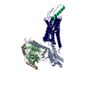 7x9bC 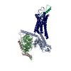 7x9cC M: map data used to model this data C: citing same article ( |
|---|---|
| Similar structure data |

-Links
 PDBj
PDBj


- Assembly
| Deposited unit | 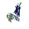
|
|---|---|
| 1 |
|
-Components
| #1: Protein | Mass: 43462.648 Da / Num. of mol.: 1 Source method: isolated from a genetically manipulated source Source: (gene. exp.) Homo sapiens (human) / Gene: NPY1R, NPYR, NPYY1 / Production host:  Trichoplusia ni (cabbage looper) / References: UniProt: P25929 Trichoplusia ni (cabbage looper) / References: UniProt: P25929 |
|---|---|
| #2: Protein | Mass: 40414.047 Da / Num. of mol.: 1 / Mutation: S47N, G203A, E245A, A326S Source method: isolated from a genetically manipulated source Source: (gene. exp.) Homo sapiens (human) / Gene: GNAI1 / Production host: Trichoplusia ni (cabbage looper) / References: UniProt: P63096 |
| #3: Protein | Mass: 38744.371 Da / Num. of mol.: 1 Source method: isolated from a genetically manipulated source Source: (gene. exp.) Homo sapiens (human) / Gene: GNB1 / Production host: Trichoplusia ni (cabbage looper) / References: UniProt: P62873 |
| #4: Protein | Mass: 7861.143 Da / Num. of mol.: 1 Source method: isolated from a genetically manipulated source Source: (gene. exp.) Homo sapiens (human) / Production host: Trichoplusia ni (cabbage looper) |
| #5: Protein/peptide | Mass: 4277.735 Da / Num. of mol.: 1 / Source method: obtained synthetically / Source: (synth.) Homo sapiens (human) / References: UniProt: P01303 |
| Has ligand of interest | N |
-Experimental details
-Experiment
| Experiment | Method: ELECTRON MICROSCOPY |
|---|---|
| EM experiment | Aggregation state: PARTICLE / 3D reconstruction method: single particle reconstruction |
- Sample preparation
| Component |
| ||||||||||||||||||||||||
|---|---|---|---|---|---|---|---|---|---|---|---|---|---|---|---|---|---|---|---|---|---|---|---|---|---|
| Source (natural) | Organism: Homo sapiens (human) | ||||||||||||||||||||||||
| Source (recombinant) | Organism: Trichoplusia ni (cabbage looper) | ||||||||||||||||||||||||
| Buffer solution | pH: 7.5 | ||||||||||||||||||||||||
| Specimen | Embedding applied: NO / Shadowing applied: NO / Staining applied: NO / Vitrification applied: YES | ||||||||||||||||||||||||
| Vitrification | Cryogen name: ETHANE |
- Electron microscopy imaging
| Experimental equipment |  Model: Titan Krios / Image courtesy: FEI Company |
|---|---|
| Microscopy | Model: FEI TITAN KRIOS |
| Electron gun | Electron source:  FIELD EMISSION GUN / Accelerating voltage: 300 kV / Illumination mode: FLOOD BEAM FIELD EMISSION GUN / Accelerating voltage: 300 kV / Illumination mode: FLOOD BEAM |
| Electron lens | Mode: BRIGHT FIELD / Nominal defocus max: 2300 nm / Nominal defocus min: 1300 nm |
| Image recording | Electron dose: 70 e/Å2 / Film or detector model: GATAN K3 BIOQUANTUM (6k x 4k) |
- Processing
| Software |
| ||||||||||||||||||||||||
|---|---|---|---|---|---|---|---|---|---|---|---|---|---|---|---|---|---|---|---|---|---|---|---|---|---|
| CTF correction | Type: NONE | ||||||||||||||||||||||||
| 3D reconstruction | Resolution: 3.2 Å / Resolution method: FSC 0.143 CUT-OFF / Num. of particles: 423400 / Symmetry type: POINT | ||||||||||||||||||||||||
| Refinement | Cross valid method: NONE Stereochemistry target values: GeoStd + Monomer Library + CDL v1.2 | ||||||||||||||||||||||||
| Displacement parameters | Biso mean: 92.41 Å2 | ||||||||||||||||||||||||
| Refine LS restraints |
|
