 ムービー
ムービー コントローラー


+ データを開く

- 基本情報
基本情報
| 登録情報 | データベース: PDB / ID: 7x7m | ||||||
|---|---|---|---|---|---|---|---|
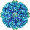
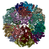
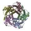
| タイトル | Lumazine Synthase from Aquifex aeolicus | ||||||
 要素 要素 | 6,7-dimethyl-8-ribityllumazine synthase | ||||||
 キーワード キーワード | TRANSFERASE / Thermostable Transferase | ||||||
| 機能・相同性 |  機能・相同性情報 機能・相同性情報6,7-dimethyl-8-ribityllumazine synthase / 6,7-dimethyl-8-ribityllumazine synthase activity / riboflavin synthase complex / riboflavin biosynthetic process / cytosol 類似検索 - 分子機能 | ||||||
| 生物種 |   Aquifex aeolicus VF5 (バクテリア) Aquifex aeolicus VF5 (バクテリア) | ||||||
| 手法 | 電子顕微鏡法 / 単粒子再構成法 / クライオ電子顕微鏡法 / 解像度: 2.33 Å | ||||||
 データ登録者 データ登録者 | Sobhy, M.A. / Hamdan, S.M. | ||||||
| 資金援助 |  サウジアラビア, 1件 サウジアラビア, 1件
| ||||||
 引用 引用 | ジャーナル: PLoS One / 年: 2022 タイトル: Cryo-electron structures of the extreme thermostable enzymes Sulfur Oxygenase Reductase and Lumazine Synthase. 著者: Mohamed A Sobhy / Lingyun Zhao / Dalaver Anjum / Ali Behzad / Masateru Takahashi / Muhammad Tehseen / Alfredo De Biasio / Rachid Sougrat / Samir Hamdan / 要旨: Thermostable enzymes have the potential for use in a wide variety of biotechnological applications. Cryo-electron microscopy (cryo-EM) enables the imaging of biomolecules in their native aqueous ...Thermostable enzymes have the potential for use in a wide variety of biotechnological applications. Cryo-electron microscopy (cryo-EM) enables the imaging of biomolecules in their native aqueous environment. Here, we present high resolution cryo-EM structures of two thermostable enzymes that exhibit multimeric cage-like structures arranged into two different point-group symmetries. First, we determined the structure of the Sulfur Oxygenase Reductase (SOR) enzyme that catalyzes both the oxygenation and disproportionation of elemental sulfur in Archea and is composed of 24 homomeric units each of MW ≃ 35 kDa arranged in octahedral symmetry. The structure of SOR from Acidianus ambivalens (7X9W) was determined at 2.78 Å resolution. The active site of each subunit inside the central nanocompartment is composed of Fe3+ coordinated to two water molecules and the three amino acids (H86, H90 and E114). Second, we determined the structure of Lumazine Synthase (LS) from Aquifex aeolicus (7X7M) at 2.33 Å resolution. LS forms a cage-like structure consisting of 60 identical subunits each of MW ≃ 15 kDa arranged in a strict icosahedral symmetry. The LS subunits are interconnected by ion-pair network. Due to their thermostability and relatively easy purification scheme, both SOR and LS can serve as a model for the catalytic and structural characterization of biocatalysts as well as a benchmark for cryo-EM sample preparation, optimization of the acquisition parameters and 3D reconstruction. | ||||||
| 履歴 |
|
- 構造の表示
構造の表示
| 構造ビューア | 分子: MolmilJmol/JSmol |
|---|
- ダウンロードとリンク
-ダウンロード
| PDBx/mmCIF形式 | 7x7m.cif.gz | 1.4 MB | 表示 | PDBx/mmCIF形式 |
|---|---|---|---|---|
| PDB形式 | pdb7x7m.ent.gz | 1.2 MB | 表示 | PDB形式 |
| PDBx/mmJSON形式 | 7x7m.json.gz | ツリー表示 | PDBx/mmJSON形式 | |
| その他 |  その他のダウンロード その他のダウンロード |
-検証レポート
| 文書・要旨 | 7x7m_validation.pdf.gz | 1.4 MB | 表示 | wwPDB検証レポート |
|---|---|---|---|---|
| 文書・詳細版 | 7x7m_full_validation.pdf.gz | 1.4 MB | 表示 | |
| XML形式データ | 7x7m_validation.xml.gz | 192.2 KB | 表示 | |
| CIF形式データ | 7x7m_validation.cif.gz | 263.5 KB | 表示 | |
| アーカイブディレクトリ | https://data.pdbj.org/pub/pdb/validation_reports/x7/7x7mftp://data.pdbj.org/pub/pdb/validation_reports/x7/7x7m | HTTPS FTP |
-関連構造データ

-リンク
 PDBj
PDBj- 集合体
集合体
| 登録構造単位 | 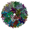
|
|---|---|
| 1 |
|
-要素
| #1: タンパク質 | 分子量: 16727.201 Da / 分子数: 60 / 由来タイプ: 組換発現 由来: (組換発現) Aquifex aeolicus VF5 (バクテリア)株: VF5 / 遺伝子: ribH / 発現宿主: 参照: UniProt: O66529, 6,7-dimethyl-8-ribityllumazine synthase |
|---|
-実験情報
-実験
| 実験 | 手法: 電子顕微鏡法 |
|---|---|
| EM実験 | 試料の集合状態: PARTICLE / 3次元再構成法: 単粒子再構成法 |
- 試料調製
試料調製
| 構成要素 | 名称: CryoEM structure of Lumazine synthase composed of 60 identical subunits タイプ: COMPLEX / Entity ID: all / 由来: RECOMBINANT | |||||||||||||||
|---|---|---|---|---|---|---|---|---|---|---|---|---|---|---|---|---|
| 由来(天然) | 生物種: Aquifex aeolicus (バクテリア) | |||||||||||||||
| 由来(組換発現) | 生物種: | |||||||||||||||
| 緩衝液 | pH: 7.5 | |||||||||||||||
| 緩衝液成分 |
| |||||||||||||||
| 試料 | 濃度: 6 mg/ml / 包埋: NO / シャドウイング: NO / 染色: NO / 凍結: YES | |||||||||||||||
| 試料支持 | グリッドの材料: GOLD / グリッドのサイズ: 300 divisions/in. / グリッドのタイプ: Quantifoil R2/2 | |||||||||||||||
| 急速凍結 | 装置: FEI VITROBOT MARK I / 凍結剤: ETHANE / 湿度: 100 % / 凍結前の試料温度: 295 K 詳細: The purified protein was applied to the glow discharged grids inside the chamber, blotted for a duration of 1.5 s, and then plunge-frozen in liquid ethane cooled by liquid nitrogen. |
- 電子顕微鏡撮影
| 実験機器 |  モデル: Titan Krios / 画像提供: FEI Company |
|---|---|
| 顕微鏡 | モデル: FEI TITAN KRIOS |
| 電子銃 | 電子線源:  FIELD EMISSION GUN / 加速電圧: 300 kV / 照射モード: SPOT SCAN FIELD EMISSION GUN / 加速電圧: 300 kV / 照射モード: SPOT SCAN |
| 電子レンズ | モード: BRIGHT FIELD / 倍率(公称値): 130000 X / 最大 デフォーカス(公称値): 1200 nm / 最小 デフォーカス(公称値): 200 nm |
| 試料ホルダ | 試料ホルダーモデル: FEI TITAN KRIOS AUTOGRID HOLDER |
| 撮影 | 電子線照射量: 70 e/Å2 / 検出モード: SUPER-RESOLUTION フィルム・検出器のモデル: GATAN K2 SUMMIT (4k x 4k) |
| 画像スキャン | 動画フレーム数/画像: 50 |
- 解析
解析
| ソフトウェア | 名称: PHENIX / バージョン: 1.18.2_3874: / 分類: 精密化 | ||||||||||||||||||||||||
|---|---|---|---|---|---|---|---|---|---|---|---|---|---|---|---|---|---|---|---|---|---|---|---|---|---|
| EMソフトウェア | 名称: RELION / カテゴリ: 3次元再構成 | ||||||||||||||||||||||||
| CTF補正 | タイプ: NONE | ||||||||||||||||||||||||
| 粒子像の選択 | 選択した粒子像数: 344110 詳細: particles were selected by LoG picking from 1516 micrographs | ||||||||||||||||||||||||
| 対称性 | 点対称性: I (正20面体型対称) | ||||||||||||||||||||||||
| 3次元再構成 | 解像度: 2.33 Å / 解像度の算出法: FSC 0.143 CUT-OFF / 粒子像の数: 103328 / クラス平均像の数: 1 / 対称性のタイプ: POINT | ||||||||||||||||||||||||
| 拘束条件 |
|
