 Movie
Movie Controller


+ Open data

- Basic information
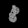
| Entry | Database: PDB / ID: 7x3v | ||||||
|---|---|---|---|---|---|---|---|
| Title | Cryo-EM structure of IOC3-N2 nucleosome | ||||||
 Components Components |
| ||||||
 Keywords Keywords | PROTEIN BINDING/DNA / PROTEIN BINDING-DNA COMPLEX | ||||||
| Function / homology |  Function and homology information Function and homology informationIsw1a complex / Isw1 complex / sister chromatid cohesion / structural constituent of chromatin / nucleosome / nucleosome assembly / heterochromatin formation / chromatin remodeling / protein heterodimerization activity / regulation of DNA-templated transcription ...Isw1a complex / Isw1 complex / sister chromatid cohesion / structural constituent of chromatin / nucleosome / nucleosome assembly / heterochromatin formation / chromatin remodeling / protein heterodimerization activity / regulation of DNA-templated transcription / DNA binding / nucleus Similarity search - Function | ||||||
| Biological species |  synthetic construct (others) | ||||||
| Method | ELECTRON MICROSCOPY / single particle reconstruction / cryo EM / Resolution: 3.09 Å | ||||||
 Authors Authors | Lifei, L. / Kangjing, C. / Chen, Z. | ||||||
| Funding support |  China, 1items China, 1items
| ||||||
 Citation Citation | Journal: Nat Struct Mol Biol / Year: 2024 Title: Structure of the ISW1a complex bound to the dinucleosome. Authors: Lifei Li / Kangjing Chen / Youyang Sia / Pengjing Hu / Youpi Ye / Zhucheng Chen / Abstract: Nucleosomes are basic repeating units of chromatin and form regularly spaced arrays in cells. Chromatin remodelers alter the positions of nucleosomes and are vital in regulating chromatin ...Nucleosomes are basic repeating units of chromatin and form regularly spaced arrays in cells. Chromatin remodelers alter the positions of nucleosomes and are vital in regulating chromatin organization and gene expression. Here we report the cryo-EM structure of chromatin remodeler ISW1a complex from Saccharomyces cerevisiae bound to the dinucleosome. Each subunit of the complex recognizes a different nucleosome. The motor subunit binds to the mobile nucleosome and recognizes the acidic patch through two arginine residues, while the DNA-binding module interacts with the entry DNA at the nucleosome edge. This nucleosome-binding mode provides the structural basis for linker DNA sensing of the motor. Notably, the Ioc3 subunit recognizes the disk face of the adjacent nucleosome through interacting with the H4 tail, the acidic patch and the nucleosomal DNA, which plays a role in the spacing activity in vitro and in nucleosome organization and cell fitness in vivo. Together, these findings support the nucleosome spacing activity of ISW1a and add a new mode of nucleosome remodeling in the context of a chromatin environment. | ||||||
| History |
|
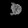
- Structure visualization
| Structure viewer | Molecule: MolmilJmol/JSmol |
|---|
- Downloads & links
-Download
| PDBx/mmCIF format | 7x3v.cif.gz | 484.1 KB | Display | PDBx/mmCIF format |
|---|---|---|---|---|
| PDB format | pdb7x3v.ent.gz | 355.9 KB | Display | PDB format |
| PDBx/mmJSON format | 7x3v.json.gz | Tree view | PDBx/mmJSON format | |
| Others |  Other downloads Other downloads |
-Validation report
| Arichive directory | https://data.pdbj.org/pub/pdb/validation_reports/x3/7x3vftp://data.pdbj.org/pub/pdb/validation_reports/x3/7x3v | HTTPS FTP |
|---|
-Related structure data
| Related structure data | 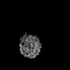 32994MC 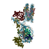 7x3tC 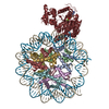 7x3wC 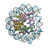 7x3xC M: map data used to model this data C: citing same article ( |
|---|---|
| Similar structure data |

-Links
 PDBj
PDBj


- Assembly
| Deposited unit | 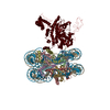
|
|---|---|
| 1 |
|
-Components
-Protein , 5 types, 9 molecules AEBFCGDHU
| #1: Protein | Mass: 15435.126 Da / Num. of mol.: 2 Source method: isolated from a genetically manipulated source Source: (gene. exp.)  #2: Protein | Mass: 11394.426 Da / Num. of mol.: 2 Source method: isolated from a genetically manipulated source Source: (gene. exp.) #3: Protein | Mass: 14109.436 Da / Num. of mol.: 2 Source method: isolated from a genetically manipulated source Source: (gene. exp.) #4: Protein | Mass: 13965.265 Da / Num. of mol.: 2 Source method: isolated from a genetically manipulated source Source: (gene. exp.) #7: Protein | | Mass: 72422.539 Da / Num. of mol.: 1 Source method: isolated from a genetically manipulated source Source: (gene. exp.) |
|---|
-DNA chain , 2 types, 2 molecules IJ
| #5: DNA chain | Mass: 45154.770 Da / Num. of mol.: 1 / Source method: obtained synthetically / Source: (synth.) synthetic construct (others) |
|---|---|
| #6: DNA chain | Mass: 45265.824 Da / Num. of mol.: 1 / Source method: obtained synthetically / Source: (synth.) synthetic construct (others) |
-Experimental details
-Experiment
| Experiment | Method: ELECTRON MICROSCOPY |
|---|---|
| EM experiment | Aggregation state: PARTICLE / 3D reconstruction method: single particle reconstruction |
- Sample preparation
| Component |
| ||||||||||||||||||||||||||||||
|---|---|---|---|---|---|---|---|---|---|---|---|---|---|---|---|---|---|---|---|---|---|---|---|---|---|---|---|---|---|---|---|
| Molecular weight | Value: 392 kDa/nm / Experimental value: YES | ||||||||||||||||||||||||||||||
| Source (natural) |
| ||||||||||||||||||||||||||||||
| Source (recombinant) |
| ||||||||||||||||||||||||||||||
| Buffer solution | pH: 7.5 | ||||||||||||||||||||||||||||||
| Buffer component |
| ||||||||||||||||||||||||||||||
| Specimen | Conc.: 1 mg/ml / Embedding applied: NO / Shadowing applied: NO / Staining applied: NO / Vitrification applied: YES | ||||||||||||||||||||||||||||||
| Vitrification | Cryogen name: ETHANE |
- Electron microscopy imaging
| Experimental equipment |  Model: Titan Krios / Image courtesy: FEI Company |
|---|---|
| Microscopy | Model: FEI TITAN KRIOS |
| Electron gun | Electron source:  FIELD EMISSION GUN / Accelerating voltage: 300 kV / Illumination mode: OTHER FIELD EMISSION GUN / Accelerating voltage: 300 kV / Illumination mode: OTHER |
| Electron lens | Mode: BRIGHT FIELD / Nominal defocus max: 1800 nm / Nominal defocus min: 1400 nm |
| Image recording | Electron dose: 50 e/Å2 / Film or detector model: GATAN K3 (6k x 4k) |
- Processing
| CTF correction | Type: NONE | ||||||||||||||||||||||||
|---|---|---|---|---|---|---|---|---|---|---|---|---|---|---|---|---|---|---|---|---|---|---|---|---|---|
| 3D reconstruction | Resolution: 3.09 Å / Resolution method: FSC 0.143 CUT-OFF / Num. of particles: 212026 / Algorithm: FOURIER SPACE / Symmetry type: POINT | ||||||||||||||||||||||||
| Refinement | Cross valid method: NONE Stereochemistry target values: GeoStd + Monomer Library + CDL v1.2 | ||||||||||||||||||||||||
| Displacement parameters | Biso mean: 103.64 Å2 | ||||||||||||||||||||||||
| Refine LS restraints |
|
