 Movie
Movie Controller

[English] 日本語
 Yorodumi
- PDB-7woo: Cryo-EM structure of the inner ring protomer of the Saccharomyces... -
+ Open data

- Basic information
| Entry | Database: PDB / ID: 7woo | ||||||
|---|---|---|---|---|---|---|---|
| Title | Cryo-EM structure of the inner ring protomer of the Saccharomyces cerevisiae nuclear pore complex | ||||||
 Components Components |
| ||||||
 Keywords Keywords | TRANSPORT PROTEIN / nuclear pore complex / inner ring / protomer / Saccharomyces cerevisiae | ||||||
| Function / homology |  Function and homology information Function and homology informationnuclear pore linkers / mRNA export from nucleus in response to heat stress / chromosome, subtelomeric region / nuclear pore inner ring / nuclear pore central transport channel / transcription-dependent tethering of RNA polymerase II gene DNA at nuclear periphery / regulation of nucleocytoplasmic transport / telomere tethering at nuclear periphery / nuclear pore complex assembly / protein localization to nuclear inner membrane ...nuclear pore linkers / mRNA export from nucleus in response to heat stress / chromosome, subtelomeric region / nuclear pore inner ring / nuclear pore central transport channel / transcription-dependent tethering of RNA polymerase II gene DNA at nuclear periphery / regulation of nucleocytoplasmic transport / telomere tethering at nuclear periphery / nuclear pore complex assembly / protein localization to nuclear inner membrane / nuclear pore organization / Regulation of Glucokinase by Glucokinase Regulatory Protein / : / nuclear pore cytoplasmic filaments / Regulation of HSF1-mediated heat shock response / post-transcriptional tethering of RNA polymerase II gene DNA at nuclear periphery / nuclear pore nuclear basket / tRNA export from nucleus / structural constituent of nuclear pore / SUMOylation of SUMOylation proteins / SUMOylation of RNA binding proteins / RNA export from nucleus / SUMOylation of chromatin organization proteins / nuclear localization sequence binding / nucleocytoplasmic transport / NLS-bearing protein import into nucleus / poly(A)+ mRNA export from nucleus / ribosomal large subunit export from nucleus / nuclear pore / mRNA transport / ribosomal small subunit export from nucleus / nuclear periphery / molecular condensate scaffold activity / promoter-specific chromatin binding / chromosome segregation / phospholipid binding / protein import into nucleus / nuclear envelope / protein transport / heterochromatin formation / nuclear membrane / amyloid fibril formation / chromatin binding / protein-containing complex binding / DNA binding / RNA binding / identical protein binding / nucleus Similarity search - Function | ||||||
| Biological species |  | ||||||
| Method | ELECTRON MICROSCOPY / single particle reconstruction / cryo EM / Resolution: 3.71 Å | ||||||
 Authors Authors | Li, Z.Q. / Chen, S.J.B. / Zhao, L. / Sui, S.F. | ||||||
| Funding support |  China, 1items China, 1items
| ||||||
 Citation Citation | Journal: Cell Res / Year: 2022 Title: Near-atomic structure of the inner ring of the Saccharomyces cerevisiae nuclear pore complex. Authors: Zongqiang Li / Shuaijiabin Chen / Liang Zhao / Guoqiang Huang / Xiong Pi / Shan Sun / Peiyi Wang / Sen-Fang Sui / Abstract: Nuclear pore complexes (NPCs) mediate bidirectional nucleocytoplasmic transport of substances in eukaryotic cells. However, the accurate molecular arrangement of NPCs remains enigmatic owing to their ...Nuclear pore complexes (NPCs) mediate bidirectional nucleocytoplasmic transport of substances in eukaryotic cells. However, the accurate molecular arrangement of NPCs remains enigmatic owing to their huge size and highly dynamic nature. Here we determined the structure of the asymmetric unit of the inner ring (IR monomer) at 3.73 Å resolution by single-particle cryo-electron microscopy, and created an atomic model of the intact IR consisting of 192 molecules of 8 nucleoporins. In each IR monomer, the Z-shaped Nup188-Nup192 complex in the middle layer is sandwiched by two approximately parallel rhomboidal structures in the inner and outer layers, while Nup188, Nup192 and Nic96 link all subunits to constitute a relatively stable IR monomer. In contrast, the intact IR is assembled by loose and instable interactions between IR monomers. These structures, together with previously reported structural information of IR, reveal two distinct interaction modes between IR monomers and extensive flexible connections in IR assembly, providing a structural basis for the stability and malleability of IR. | ||||||
| History |
|
- Structure visualization
| Structure viewer | Molecule: MolmilJmol/JSmol |
|---|
- Downloads & links
-Download
| PDBx/mmCIF format | 7woo.cif.gz | 1.5 MB | Display | PDBx/mmCIF format |
|---|---|---|---|---|
| PDB format | pdb7woo.ent.gz | 1.2 MB | Display | PDB format |
| PDBx/mmJSON format | 7woo.json.gz | Tree view | PDBx/mmJSON format | |
| Others |  Other downloads Other downloads |
-Validation report
| Arichive directory | https://data.pdbj.org/pub/pdb/validation_reports/wo/7wooftp://data.pdbj.org/pub/pdb/validation_reports/wo/7woo | HTTPS FTP |
|---|
-Related structure data
| Related structure data |  32653MC 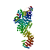 7wo9C  7wotC M: map data used to model this data C: citing same article ( |
|---|---|
| Similar structure data |


-Links
 PDBj
PDBj

- Assembly
| Deposited unit | 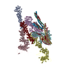
|
|---|---|
| 1 |
|
-Components
-Protein , 8 types, 12 molecules AZCDEFGJHKIL
| #1: Protein | Mass: 96291.586 Da / Num. of mol.: 2 / Source method: isolated from a natural source / Source: (natural) #2: Protein | | Mass: 156827.484 Da / Num. of mol.: 1 / Source method: isolated from a natural source / Source: (natural) #3: Protein | | Mass: 169651.969 Da / Num. of mol.: 1 / Source method: isolated from a natural source / Source: (natural) #4: Protein | | Mass: 188753.281 Da / Num. of mol.: 1 / Source method: isolated from a natural source / Source: (natural) #5: Protein | | Mass: 191718.125 Da / Num. of mol.: 1 / Source method: isolated from a natural source / Source: (natural) #6: Protein | Mass: 49174.762 Da / Num. of mol.: 2 / Source method: isolated from a natural source / Source: (natural) #7: Protein | Mass: 57547.145 Da / Num. of mol.: 2 / Source method: isolated from a natural source / Source: (natural) #8: Protein | Mass: 86611.672 Da / Num. of mol.: 2 / Source method: isolated from a natural source / Source: (natural) |
|---|
-Experimental details
-Experiment
| Experiment | Method: ELECTRON MICROSCOPY |
|---|---|
| EM experiment | Aggregation state: PARTICLE / 3D reconstruction method: single particle reconstruction |
- Sample preparation
| Component | Name: Cryo-EM structure of the inner ring protomer of the Saccharomyces cerevisiae nuclear pore complex Type: COMPLEX / Entity ID: #4 / Source: NATURAL |
|---|---|
| Source (natural) | Organism: |
| Buffer solution | pH: 7.5 |
| Specimen | Embedding applied: NO / Shadowing applied: NO / Staining applied: NO / Vitrification applied: YES |
| Vitrification | Cryogen name: ETHANE |
- Electron microscopy imaging
| Experimental equipment |  Model: Titan Krios / Image courtesy: FEI Company |
|---|---|
| Microscopy | Model: FEI TITAN KRIOS |
| Electron gun | Electron source:  FIELD EMISSION GUN / Accelerating voltage: 300 kV / Illumination mode: FLOOD BEAM FIELD EMISSION GUN / Accelerating voltage: 300 kV / Illumination mode: FLOOD BEAM |
| Electron lens | Mode: BRIGHT FIELD / Nominal defocus max: 3000 nm / Nominal defocus min: 1500 nm |
| Image recording | Electron dose: 50 e/Å2 / Film or detector model: GATAN K3 BIOQUANTUM (6k x 4k) |
- Processing
| CTF correction | Type: NONE |
|---|---|
| 3D reconstruction | Resolution: 3.71 Å / Resolution method: FSC 0.143 CUT-OFF / Num. of particles: 1266268 / Symmetry type: POINT |
