 Movie
Movie Controller


+ Open data

- Basic information
| Entry | Database: PDB / ID: 7wb5 | ||||||
|---|---|---|---|---|---|---|---|
| Title | local structure of hu33 and spike | ||||||
 Components Components |
| ||||||
 Keywords Keywords | VIRAL PROTEIN / complex / spike / antibody | ||||||
| Function / homology | Betacoronavirus spike (S) glycoprotein S1 subunit C-terminal (CTD) domain profile. / Spike (S) protein S1 subunit, receptor-binding domain, betacoronavirus / Spike S1 subunit, receptor binding domain superfamily, betacoronavirus / Betacoronavirus spike glycoprotein S1, receptor binding / Coronavirus spike glycoprotein S1, C-terminal / Coronavirus spike glycoprotein S1, C-terminal / Surface glycoprotein Function and homology information Function and homology information | ||||||
| Biological species |  Homo sapiens (human) Homo sapiens (human)  Severe acute respiratory syndrome coronavirus 2 Severe acute respiratory syndrome coronavirus 2 | ||||||
| Method | ELECTRON MICROSCOPY / single particle reconstruction / cryo EM / Resolution: 3.7 Å | ||||||
 Authors Authors | Pulan, L. | ||||||
| Funding support |  China, 1items China, 1items
| ||||||
 Citation Citation | Journal: Signal Transduct Target Ther / Year: 2022 Title: A non-ACE2-blocking neutralizing antibody against Omicron-included SARS-CoV-2 variants. Authors: Xiaomin Duan / Rui Shi / Pulan Liu / Qingrui Huang / Fengze Wang / Xinyu Chen / Hui Feng / Weijin Huang / Junyu Xiao / Jinghua Yan / | ||||||
| History |
|
- Structure visualization
| Structure viewer | Molecule: MolmilJmol/JSmol |
|---|
- Downloads & links
-Download
| PDBx/mmCIF format | 7wb5.cif.gz | 81.6 KB | Display | PDBx/mmCIF format |
|---|---|---|---|---|
| PDB format | pdb7wb5.ent.gz | 63.6 KB | Display | PDB format |
| PDBx/mmJSON format | 7wb5.json.gz | Tree view | PDBx/mmJSON format | |
| Others |  Other downloads Other downloads |
-Validation report
| Summary document | 7wb5_validation.pdf.gz | 823.8 KB | Display | wwPDB validaton report |
|---|---|---|---|---|
| Full document | 7wb5_full_validation.pdf.gz | 836.3 KB | Display | |
| Data in XML | 7wb5_validation.xml.gz | 21.8 KB | Display | |
| Data in CIF | 7wb5_validation.cif.gz | 30.3 KB | Display | |
| Arichive directory | https://data.pdbj.org/pub/pdb/validation_reports/wb/7wb5ftp://data.pdbj.org/pub/pdb/validation_reports/wb/7wb5 | HTTPS FTP |
-Related structure data
| Related structure data |  32395MC  7wbhC M: map data used to model this data C: citing same article ( |
|---|---|
| Similar structure data |

-Links
 PDBj
PDBj

- Assembly
| Deposited unit | 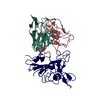
|
|---|---|
| 1 |
|
-Components
| #1: Protein | Mass: 13162.599 Da / Num. of mol.: 1 Source method: isolated from a genetically manipulated source Source: (gene. exp.) Homo sapiens (human) / Production host: Homo sapiens (human) |
|---|---|
| #2: Antibody | Mass: 11717.014 Da / Num. of mol.: 1 Source method: isolated from a genetically manipulated source Source: (gene. exp.) Homo sapiens (human) / Production host: Homo sapiens (human) |
| #3: Protein | Mass: 20745.172 Da / Num. of mol.: 1 Source method: isolated from a genetically manipulated source Source: (gene. exp.) Severe acute respiratory syndrome coronavirus 2Production host: Homo sapiens (human) / References: UniProt: A0A7U3CI26 |
| #4: Polysaccharide | beta-D-mannopyranose-(1-4)-2-acetamido-2-deoxy-beta-D-glucopyranose-(1-4)-2-acetamido-2-deoxy-beta- ...beta-D-mannopyranose-(1-4)-2-acetamido-2-deoxy-beta-D-glucopyranose-(1-4)-2-acetamido-2-deoxy-beta-D-glucopyranose Source method: isolated from a genetically manipulated source |
| Has ligand of interest | N |
-Experimental details
-Experiment
| Experiment | Method: ELECTRON MICROSCOPY |
|---|---|
| EM experiment | Aggregation state: PARTICLE / 3D reconstruction method: single particle reconstruction |
- Sample preparation
| Component | Name: local structure of R33 and RBD / Type: COMPLEX / Entity ID: #1-#3 / Source: RECOMBINANT |
|---|---|
| Source (natural) | Organism: Homo sapiens (human) |
| Source (recombinant) | Organism: Homo sapiens (human) |
| Buffer solution | pH: 7.2 |
| Specimen | Embedding applied: NO / Shadowing applied: NO / Staining applied: NO / Vitrification applied: YES |
| Vitrification | Cryogen name: ETHANE |
- Electron microscopy imaging
| Experimental equipment |  Model: Titan Krios / Image courtesy: FEI Company |
|---|---|
| Microscopy | Model: FEI TITAN KRIOS |
| Electron gun | Electron source:  FIELD EMISSION GUN / Accelerating voltage: 300 kV / Illumination mode: SPOT SCAN FIELD EMISSION GUN / Accelerating voltage: 300 kV / Illumination mode: SPOT SCAN |
| Electron lens | Mode: BRIGHT FIELD / Nominal defocus max: 1500 nm / Nominal defocus min: 1000 nm |
| Image recording | Electron dose: 50 e/Å2 / Film or detector model: GATAN K3 BIOQUANTUM (6k x 4k) |
- Processing
| CTF correction | Type: PHASE FLIPPING AND AMPLITUDE CORRECTION |
|---|---|
| 3D reconstruction | Resolution: 3.7 Å / Resolution method: FSC 0.143 CUT-OFF / Num. of particles: 222776 / Symmetry type: POINT |
