 Movie
Movie Controller

[English] 日本語
 Yorodumi
- PDB-7wa1: Structure of SARS-CoV-2 spike receptor-binding domain F486L mutat... -
+ Open data

- Basic information
| Entry | Database: PDB / ID: 7wa1 | ||||||
|---|---|---|---|---|---|---|---|
| Title | Structure of SARS-CoV-2 spike receptor-binding domain F486L mutation complexed with American mink ACE2 | ||||||
 Components Components |
| ||||||
 Keywords Keywords | VIRAL PROTEIN | ||||||
| Function / homology |  Function and homology information Function and homology informationsymbiont-mediated disruption of host tissue / Maturation of spike protein / Translation of Structural Proteins / Virion Assembly and Release / host cell surface / host extracellular region / symbiont-mediated-mediated suppression of host tetherin activity / Induction of Cell-Cell Fusion / structural constituent of virion / positive regulation of viral entry into host cell ...symbiont-mediated disruption of host tissue / Maturation of spike protein / Translation of Structural Proteins / Virion Assembly and Release / host cell surface / host extracellular region / symbiont-mediated-mediated suppression of host tetherin activity / Induction of Cell-Cell Fusion / structural constituent of virion / positive regulation of viral entry into host cell / membrane fusion / host cell endoplasmic reticulum-Golgi intermediate compartment membrane / Attachment and Entry / entry receptor-mediated virion attachment to host cell / receptor-mediated virion attachment to host cell / host cell surface receptor binding / symbiont-mediated suppression of host innate immune response / endocytosis involved in viral entry into host cell / receptor ligand activity / fusion of virus membrane with host plasma membrane / fusion of virus membrane with host endosome membrane / viral envelope / symbiont entry into host cell / virion attachment to host cell / host cell plasma membrane / SARS-CoV-2 activates/modulates innate and adaptive immune responses / virion membrane / membrane / identical protein binding / plasma membrane Similarity search - Function | ||||||
| Biological species |  Homo sapiens (human) Homo sapiens (human)  Severe acute respiratory syndrome coronavirus 2 Severe acute respiratory syndrome coronavirus 2 | ||||||
| Method | ELECTRON MICROSCOPY / single particle reconstruction / cryo EM / Resolution: 2.9 Å | ||||||
 Authors Authors | Su, C. / Qi, J.X. / Gao, G.F. | ||||||
| Funding support | 1items
| ||||||
 Citation Citation | Journal: J Virol / Year: 2022 Title: Molecular Basis of Mink ACE2 Binding to SARS-CoV-2 and Its Mink-Derived Variants. Authors: Chao Su / Juanhua He / Pengcheng Han / Bin Bai / Dedong Li / Jian Cao / Mingxiong Tian / Yu Hu / Anqi Zheng / Sheng Niu / Qian Chen / Xiaoyu Rong / Yanfang Zhang / Weiwei Li / Jianxun Qi / ...Authors: Chao Su / Juanhua He / Pengcheng Han / Bin Bai / Dedong Li / Jian Cao / Mingxiong Tian / Yu Hu / Anqi Zheng / Sheng Niu / Qian Chen / Xiaoyu Rong / Yanfang Zhang / Weiwei Li / Jianxun Qi / Xin Zhao / Mengsu Yang / Qihui Wang / George Fu Gao /  Abstract: Severe acute respiratory syndrome coronavirus 2 (SARS-CoV-2) is transmitted between humans and minks, and some mutations in the spike (S) protein, especially in the receptor-binding domain (RBD), ...Severe acute respiratory syndrome coronavirus 2 (SARS-CoV-2) is transmitted between humans and minks, and some mutations in the spike (S) protein, especially in the receptor-binding domain (RBD), have been identified in mink-derived viruses. Here, we examined binding of the mink angiotensin-converting enzyme 2 (ACE2) receptor to mink-derived and important human-originating variants, and we demonstrated that most of the RBD variants increased the binding affinities to mink ACE2 (mkACE2). Cryo-electron microscopy structures of the mkACE2-RBD Y453F (with a Y-to-F change at position 453) and mkACE2-RBD F486L complexes helped identify the key residues that facilitate changes in mkACE2 binding affinity. Additionally, the data indicated that the Y453F and F486L mutations reduced the binding affinities to some human monoclonal antibodies, and human vaccinated sera efficiently prevented infection of human cells by pseudoviruses expressing Y453F, F486L, or N501T RBD. Our findings provide an important molecular mechanism for the rapid adaptation of SARS-CoV-2 in minks and highlight the potential influence of the main mink-originating variants for humans. Severe acute respiratory syndrome coronavirus 2 (SARS-CoV-2) has a broad range of hosts. Mink-derived SARS-CoV-2 can transmit back to humans. There is an urgent need to understand the binding mechanism of mink-derived SARS-CoV-2 variants to mink receptor. In this study, we identified all mutations in the receptor-binding domain (RBD) of spike (S) protein from mink-derived SARS-CoV-2, and we demonstrated the enhanced binding affinity of mink angiotensin-converting enzyme 2 (ACE2) to most of the mink-derived RBD variants as well as important human-originating RBD variants. Cryo-electron microscopy structures revealed that the Y453F and F486L mutations enhanced the binding forces in the interaction interface. In addition, Y453F and F486L mutations reduced the binding affinities to some human monoclonal antibodies, and the SARS-CoV-2 pseudoviruses with Y453F, F486L, or N501T mutations were neutralized by human vaccinated sera. Therefore, our results provide valuable information for understanding the cross-species transmission mechanism of SARS-CoV-2. | ||||||
| History |
|
- Structure visualization
| Structure viewer | Molecule: MolmilJmol/JSmol |
|---|
- Downloads & links
-Download
| PDBx/mmCIF format | 7wa1.cif.gz | 168.9 KB | Display | PDBx/mmCIF format |
|---|---|---|---|---|
| PDB format | pdb7wa1.ent.gz | 128.1 KB | Display | PDB format |
| PDBx/mmJSON format | 7wa1.json.gz | Tree view | PDBx/mmJSON format | |
| Others |  Other downloads Other downloads |
-Validation report
| Arichive directory | https://data.pdbj.org/pub/pdb/validation_reports/wa/7wa1ftp://data.pdbj.org/pub/pdb/validation_reports/wa/7wa1 | HTTPS FTP |
|---|
-Related structure data
| Related structure data |  32379MC  7w8sC M: map data used to model this data C: citing same article ( |
|---|---|
| Similar structure data |

-Links
 PDBj
PDBj


- Assembly
| Deposited unit | 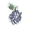
|
|---|---|
| 1 |
|
-Components
| #1: Protein | Mass: 70562.195 Da / Num. of mol.: 1 Source method: isolated from a genetically manipulated source Source: (gene. exp.) Homo sapiens (human) / Production host:  Trichoplusia ni (cabbage looper) Trichoplusia ni (cabbage looper) |
|---|---|
| #2: Protein | Mass: 25917.203 Da / Num. of mol.: 1 / Mutation: F486L Source method: isolated from a genetically manipulated source Source: (gene. exp.) Severe acute respiratory syndrome coronavirus 2Gene: S, 2 / Cell line (production host): HEK293 / Production host: Homo sapiens (human) / References: UniProt: P0DTC2 |
| #3: Chemical | ChemComp-ZN /   Mass: 65.409 Da / Num. of mol.: 1 / Source method: obtained synthetically / Formula: Zn / Feature type: SUBJECT OF INVESTIGATION Mass: 65.409 Da / Num. of mol.: 1 / Source method: obtained synthetically / Formula: Zn / Feature type: SUBJECT OF INVESTIGATION |
| Has ligand of interest | Y |
| Has protein modification | Y |
-Experimental details
-Experiment
| Experiment | Method: ELECTRON MICROSCOPY |
|---|---|
| EM experiment | Aggregation state: PARTICLE / 3D reconstruction method: single particle reconstruction |
- Sample preparation
| Component |
| ||||||||||||||||||||||||
|---|---|---|---|---|---|---|---|---|---|---|---|---|---|---|---|---|---|---|---|---|---|---|---|---|---|
| Source (natural) |
| ||||||||||||||||||||||||
| Source (recombinant) |
| ||||||||||||||||||||||||
| Buffer solution | pH: 8 | ||||||||||||||||||||||||
| Specimen | Embedding applied: NO / Shadowing applied: NO / Staining applied: NO / Vitrification applied: YES | ||||||||||||||||||||||||
| Vitrification | Cryogen name: ETHANE |
- Electron microscopy imaging
| Experimental equipment |  Model: Titan Krios / Image courtesy: FEI Company |
|---|---|
| Microscopy | Model: FEI TITAN KRIOS |
| Electron gun | Electron source:  FIELD EMISSION GUN / Accelerating voltage: 300 kV / Illumination mode: FLOOD BEAM FIELD EMISSION GUN / Accelerating voltage: 300 kV / Illumination mode: FLOOD BEAM |
| Electron lens | Mode: BRIGHT FIELD / Nominal defocus max: 2000 nm / Nominal defocus min: 1000 nm |
| Image recording | Electron dose: 50 e/Å2 / Film or detector model: FEI FALCON IV (4k x 4k) |
- Processing
| Software | Name: PHENIX / Version: 1.19.2_4158: / Classification: refinement | ||||||||||||||||||||||||
|---|---|---|---|---|---|---|---|---|---|---|---|---|---|---|---|---|---|---|---|---|---|---|---|---|---|
| CTF correction | Type: PHASE FLIPPING ONLY | ||||||||||||||||||||||||
| 3D reconstruction | Resolution: 2.9 Å / Resolution method: OTHER / Num. of particles: 464172 / Symmetry type: POINT | ||||||||||||||||||||||||
| Refinement | Cross valid method: NONE Stereochemistry target values: GeoStd + Monomer Library + CDL v1.2 | ||||||||||||||||||||||||
| Displacement parameters | Biso mean: 48.22 Å2 | ||||||||||||||||||||||||
| Refine LS restraints |
|
