 Movie
Movie Controller

[English] 日本語
 Yorodumi
- PDB-7w9p: Cryo-EM structure of human Nav1.7(E406K) in complex with auxiliar... -
+ Open data

- Basic information
| Entry | Database: PDB / ID: 7w9p | ||||||
|---|---|---|---|---|---|---|---|
| Title | Cryo-EM structure of human Nav1.7(E406K) in complex with auxiliary beta subunits, huwentoxin-IV and saxitoxin (S6IV pi helix conformer) | ||||||
 Components Components |
| ||||||
 Keywords Keywords | MEMBRANE PROTEIN / Nav1.7 / SCN9A / cryo-EM | ||||||
| Function / homology |  Function and homology information Function and homology informationresponse to pyrethroid / corticospinal neuron axon guidance / positive regulation of voltage-gated sodium channel activity / action potential propagation / detection of mechanical stimulus involved in sensory perception / voltage-gated sodium channel activity involved in cardiac muscle cell action potential / regulation of atrial cardiac muscle cell membrane depolarization / voltage-gated potassium channel activity involved in ventricular cardiac muscle cell action potential repolarization / cardiac conduction / membrane depolarization during Purkinje myocyte cell action potential ...response to pyrethroid / corticospinal neuron axon guidance / positive regulation of voltage-gated sodium channel activity / action potential propagation / detection of mechanical stimulus involved in sensory perception / voltage-gated sodium channel activity involved in cardiac muscle cell action potential / regulation of atrial cardiac muscle cell membrane depolarization / voltage-gated potassium channel activity involved in ventricular cardiac muscle cell action potential repolarization / cardiac conduction / membrane depolarization during Purkinje myocyte cell action potential / membrane depolarization during cardiac muscle cell action potential / membrane depolarization during action potential / positive regulation of sodium ion transport / regulation of sodium ion transmembrane transport / axon initial segment / regulation of ventricular cardiac muscle cell membrane repolarization / cardiac muscle cell action potential involved in contraction / sodium channel inhibitor activity / voltage-gated sodium channel complex / node of Ranvier / neuronal action potential propagation / locomotion / voltage-gated sodium channel activity / Interaction between L1 and Ankyrins / Phase 0 - rapid depolarisation / regulation of heart rate by cardiac conduction / detection of temperature stimulus involved in sensory perception of pain / behavioral response to pain / intercalated disc / sodium channel regulator activity / membrane depolarization / neuronal action potential / cardiac muscle contraction / T-tubule / sensory perception of pain / axon terminus / axon guidance / sodium ion transmembrane transport / post-embryonic development / circadian rhythm / positive regulation of neuron projection development / response to toxic substance / Sensory perception of sweet, bitter, and umami (glutamate) taste / nervous system development / response to heat / gene expression / chemical synaptic transmission / transmembrane transporter binding / perikaryon / cell adhesion / inflammatory response / axon / synapse / extracellular region / plasma membrane Similarity search - Function | ||||||
| Biological species |  Homo sapiens (human) Homo sapiens (human) | ||||||
| Method | ELECTRON MICROSCOPY / single particle reconstruction / cryo EM / Resolution: 2.9 Å | ||||||
 Authors Authors | Yan, N. / Huang, G. / Liu, D. / Wei, P. / Shen, H. | ||||||
| Funding support |  China, 1items China, 1items
| ||||||
 Citation Citation | Journal: Cell Rep / Year: 2022 Title: High-resolution structures of human Na1.7 reveal gating modulation through α-π helical transition of S6. Authors: Gaoxingyu Huang / Dongliang Liu / Weipeng Wang / Qiurong Wu / Jiaofeng Chen / Xiaojing Pan / Huaizong Shen / Nieng Yan / Abstract: Na1.7 represents a preeminent target for next-generation analgesics for its critical role in pain sensation. Here we report a 2.2-Å resolution cryo-EM structure of wild-type (WT) Na1.7 complexed ...Na1.7 represents a preeminent target for next-generation analgesics for its critical role in pain sensation. Here we report a 2.2-Å resolution cryo-EM structure of wild-type (WT) Na1.7 complexed with the β1 and β2 subunits that reveals several previously indiscernible cytosolic segments. Reprocessing of the cryo-EM data for our reported structures of Na1.7(E406K) bound to various toxins identifies two distinct conformations of S6, one composed of α helical turns only and the other containing a π helical turn in the middle. The structure of ligand-free Na1.7(E406K), determined at 3.5-Å resolution, is identical to the WT channel, confirming that binding of Huwentoxin IV or Protoxin II to VSD allosterically induces the α → π transition of S6. The local secondary structural shift leads to contraction of the intracellular gate, closure of the fenestration on the interface of repeats I and IV, and rearrangement of the binding site for the fast inactivation motif. | ||||||
| History |
|
- Structure visualization
| Structure viewer | Molecule: MolmilJmol/JSmol |
|---|
- Downloads & links
-Download
| PDBx/mmCIF format | 7w9p.cif.gz | 375.3 KB | Display | PDBx/mmCIF format |
|---|---|---|---|---|
| PDB format | pdb7w9p.ent.gz | 280.6 KB | Display | PDB format |
| PDBx/mmJSON format | 7w9p.json.gz | Tree view | PDBx/mmJSON format | |
| Others |  Other downloads Other downloads |
-Validation report
| Arichive directory | https://data.pdbj.org/pub/pdb/validation_reports/w9/7w9pftp://data.pdbj.org/pub/pdb/validation_reports/w9/7w9p | HTTPS FTP |
|---|
-Related structure data
| Related structure data | 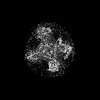 32371MC 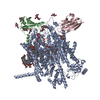 7w9kC 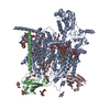 7w9lC  7w9mC 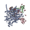 7w9tC M: map data used to model this data C: citing same article ( |
|---|---|
| Similar structure data |
-Links
 PDBj
PDBj


- Assembly
| Deposited unit | 
|
|---|---|
| 1 |
|
-Components
-Protein , 1 types, 1 molecules A
| #1: Protein | Mass: 231211.922 Da / Num. of mol.: 1 / Mutation: E406K Source method: isolated from a genetically manipulated source Source: (gene. exp.) Homo sapiens (human) / Gene: SCN9A, NENA / Production host: Homo sapiens (human) / References: UniProt: Q15858 |
|---|
-Sodium channel subunit beta- ... , 2 types, 2 molecules BC
| #2: Protein | Mass: 24732.115 Da / Num. of mol.: 1 Source method: isolated from a genetically manipulated source Source: (gene. exp.) Homo sapiens (human) / Gene: SCN1B / Production host: Homo sapiens (human) / References: UniProt: Q07699 |
|---|---|
| #3: Protein | Mass: 24355.859 Da / Num. of mol.: 1 Source method: isolated from a genetically manipulated source Source: (gene. exp.) Homo sapiens (human) / Gene: SCN2B, UNQ326/PRO386 / Production host: Homo sapiens (human) / References: UniProt: O60939 |
-Sugars , 2 types, 8 molecules 
| #4: Polysaccharide | Source method: isolated from a genetically manipulated source #6: Sugar | ChemComp-NAG /  Type: D-saccharide, beta linking / Mass: 221.208 Da / Num. of mol.: 5 / Source method: obtained synthetically / Formula: C8H15NO6 / Feature type: SUBJECT OF INVESTIGATION Type: D-saccharide, beta linking / Mass: 221.208 Da / Num. of mol.: 5 / Source method: obtained synthetically / Formula: C8H15NO6 / Feature type: SUBJECT OF INVESTIGATION |
|---|
-Non-polymers , 6 types, 29 molecules 


| #5: Chemical | ChemComp-9SL / [( Mass: 299.286 Da / Num. of mol.: 1 / Source method: obtained synthetically / Formula: C10H17N7O4 / Feature type: SUBJECT OF INVESTIGATION / Comment: toxin*YM Mass: 299.286 Da / Num. of mol.: 1 / Source method: obtained synthetically / Formula: C10H17N7O4 / Feature type: SUBJECT OF INVESTIGATION / Comment: toxin*YM | ||||||||
|---|---|---|---|---|---|---|---|---|---|
| #7: Chemical |  Mass: 792.075 Da / Num. of mol.: 3 / Source method: obtained synthetically / Formula: C42H82NO10P Mass: 792.075 Da / Num. of mol.: 3 / Source method: obtained synthetically / Formula: C42H82NO10P#8: Chemical | ChemComp-Y01 /  Mass: 486.726 Da / Num. of mol.: 6 / Source method: obtained synthetically / Formula: C31H50O4 Mass: 486.726 Da / Num. of mol.: 6 / Source method: obtained synthetically / Formula: C31H50O4#9: Chemical | ChemComp-LPE /  Mass: 510.708 Da / Num. of mol.: 13 / Source method: obtained synthetically / Formula: C26H57NO6P Mass: 510.708 Da / Num. of mol.: 13 / Source method: obtained synthetically / Formula: C26H57NO6P#10: Chemical | ChemComp-1PW / ( |  Mass: 421.508 Da / Num. of mol.: 1 / Source method: obtained synthetically / Formula: C20H40NO6P Mass: 421.508 Da / Num. of mol.: 1 / Source method: obtained synthetically / Formula: C20H40NO6P#11: Chemical | ChemComp-PCW /  Mass: 787.121 Da / Num. of mol.: 5 / Source method: obtained synthetically / Formula: C44H85NO8P / Comment: DOPC, phospholipid*YM Mass: 787.121 Da / Num. of mol.: 5 / Source method: obtained synthetically / Formula: C44H85NO8P / Comment: DOPC, phospholipid*YM |
-Details
| Has ligand of interest | Y |
|---|---|
| Has protein modification | Y |
-Experimental details
-Experiment
| Experiment | Method: ELECTRON MICROSCOPY |
|---|---|
| EM experiment | Aggregation state: PARTICLE / 3D reconstruction method: single particle reconstruction |
- Sample preparation
| Component | Name: Human voltage-gated sodium channel Nav1.7 in complex with auxiliary beta subunits Type: COMPLEX / Entity ID: #1-#3 / Source: RECOMBINANT |
|---|---|
| Molecular weight | Value: 279.99 kDa/nm / Experimental value: NO |
| Source (natural) | Organism: Homo sapiens (human) |
| Source (recombinant) | Organism: Homo sapiens (human) |
| Buffer solution | pH: 7.4 |
| Specimen | Embedding applied: NO / Shadowing applied: NO / Staining applied: NO / Vitrification applied: YES |
| Vitrification | Cryogen name: ETHANE |
- Electron microscopy imaging
| Experimental equipment |  Model: Titan Krios / Image courtesy: FEI Company |
|---|---|
| Microscopy | Model: FEI TITAN KRIOS |
| Electron gun | Electron source:  FIELD EMISSION GUN / Accelerating voltage: 300 kV / Illumination mode: FLOOD BEAM FIELD EMISSION GUN / Accelerating voltage: 300 kV / Illumination mode: FLOOD BEAM |
| Electron lens | Mode: BRIGHT FIELD / Nominal defocus max: 1800 nm / Nominal defocus min: 1500 nm |
| Image recording | Electron dose: 50 e/Å2 / Film or detector model: GATAN K3 (6k x 4k) |
- Processing
| CTF correction | Type: PHASE FLIPPING AND AMPLITUDE CORRECTION |
|---|---|
| Symmetry | Point symmetry: C1 (asymmetric) |
| 3D reconstruction | Resolution: 2.9 Å / Resolution method: FSC 0.143 CUT-OFF / Num. of particles: 194430 / Symmetry type: POINT |
