 Movie
Movie Controller

[English] 日本語
 Yorodumi
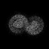
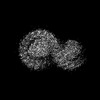
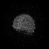
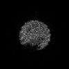
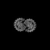
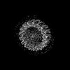
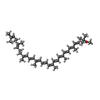
- PDB-7vor: The structure of dimeric photosynthetic RC-LH1 supercomplex in Class-1 -
+ Open data

- Basic information
| Entry | Database: PDB / ID: 7vor | ||||||||||||||||||||||||
|---|---|---|---|---|---|---|---|---|---|---|---|---|---|---|---|---|---|---|---|---|---|---|---|---|---|
| Title | The structure of dimeric photosynthetic RC-LH1 supercomplex in Class-1 | ||||||||||||||||||||||||
 Components Components |
| ||||||||||||||||||||||||
 Keywords Keywords | PHOTOSYNTHESIS / SUPERCOMPLEX / purple bacteria | ||||||||||||||||||||||||
| Function / homology |  Function and homology information Function and homology informationorganelle inner membrane / plasma membrane-derived chromatophore membrane / plasma membrane light-harvesting complex / bacteriochlorophyll binding / photosynthetic electron transport in photosystem II / photosynthesis, light reaction / photosynthesis / metal ion binding / plasma membrane Similarity search - Function | ||||||||||||||||||||||||
| Biological species |  Cereibacter sphaeroides 2.4.1 (bacteria) Cereibacter sphaeroides 2.4.1 (bacteria) | ||||||||||||||||||||||||
| Method | ELECTRON MICROSCOPY / single particle reconstruction / cryo EM / Resolution: 2.74 Å | ||||||||||||||||||||||||
 Authors Authors | Cao, P. / Li, M. / Liu, L.N. | ||||||||||||||||||||||||
| Funding support |  China, China,  United Kingdom, 7items United Kingdom, 7items
| ||||||||||||||||||||||||
 Citation Citation | Journal: Nat Commun / Year: 2022 Title: Structural basis for the assembly and quinone transport mechanisms of the dimeric photosynthetic RC-LH1 supercomplex. Authors: Peng Cao / Laura Bracun / Atsushi Yamagata / Bern M Christianson / Tatsuki Negami / Baohua Zou / Tohru Terada / Daniel P Canniffe / Mikako Shirouzu / Mei Li / Lu-Ning Liu /  Abstract: The reaction center (RC) and light-harvesting complex 1 (LH1) form a RC-LH1 core supercomplex that is vital for the primary reactions of photosynthesis in purple phototrophic bacteria. Some species ...The reaction center (RC) and light-harvesting complex 1 (LH1) form a RC-LH1 core supercomplex that is vital for the primary reactions of photosynthesis in purple phototrophic bacteria. Some species possess the dimeric RC-LH1 complex with a transmembrane polypeptide PufX, representing the largest photosynthetic complex in anoxygenic phototrophs. However, the details of the architecture and assembly mechanism of the RC-LH1 dimer are unclear. Here we report seven cryo-electron microscopy (cryo-EM) structures of RC-LH1 supercomplexes from Rhodobacter sphaeroides. Our structures reveal that two PufX polypeptides are positioned in the center of the S-shaped RC-LH1 dimer, interlocking association between the components and mediating RC-LH1 dimerization. Moreover, we identify another transmembrane peptide, designated PufY, which is located between the RC and LH1 subunits near the LH1 opening. PufY binds a quinone molecule and prevents LH1 subunits from completely encircling the RC, creating a channel for quinone/quinol exchange. Genetic mutagenesis, cryo-EM structures, and computational simulations provide a mechanistic understanding of the assembly and electron transport pathways of the RC-LH1 dimer and elucidate the roles of individual components in ensuring the structural and functional integrity of the photosynthetic supercomplex. | ||||||||||||||||||||||||
| History |
|
- Structure visualization
| Structure viewer | Molecule: MolmilJmol/JSmol |
|---|
- Downloads & links
-Download
| PDBx/mmCIF format | 7vor.cif.gz | 960 KB | Display | PDBx/mmCIF format |
|---|---|---|---|---|
| PDB format | pdb7vor.ent.gz | Display | PDB format | |
| PDBx/mmJSON format | 7vor.json.gz | Tree view | PDBx/mmJSON format | |
| Others |  Other downloads Other downloads |
-Validation report
| Arichive directory | https://data.pdbj.org/pub/pdb/validation_reports/vo/7vorftp://data.pdbj.org/pub/pdb/validation_reports/vo/7vor | HTTPS FTP |
|---|
-Related structure data
| Related structure data | 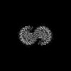 32058MC  7va9C  7vb9C  7vnmC  7vnyC  7votC  7voyC M: map data used to model this data C: citing same article ( |
|---|---|
| Similar structure data |
-Links
 PDBj
PDBj


- Assembly
| Deposited unit | 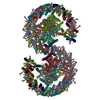
|
|---|---|
| 1 |
|
-Components
-Reaction center protein ... , 3 types, 6 molecules LlMmHh
| #1: Protein | Mass: 31477.584 Da / Num. of mol.: 2 / Source method: isolated from a natural source / Source: (natural) Cereibacter sphaeroides 2.4.1 (bacteria) / Strain: 2.4.1. / References: UniProt: Q3J1A5#2: Protein | Mass: 34529.738 Da / Num. of mol.: 2 / Source method: isolated from a natural source / Source: (natural) Cereibacter sphaeroides 2.4.1 (bacteria) / Strain: 2.4.1. / References: UniProt: Q3J1A6#3: Protein | Mass: 28066.322 Da / Num. of mol.: 2 / Source method: isolated from a natural source / Source: (natural) Cereibacter sphaeroides 2.4.1 (bacteria) / Strain: 2.4.1. / References: UniProt: Q3J170 |
|---|
-Light-harvesting protein B-875 ... , 2 types, 56 molecules ADFIKOQSUW3179adfikoqsuw5b16b9BE...
| #4: Protein | Mass: 6816.169 Da / Num. of mol.: 28 / Source method: isolated from a natural source / Source: (natural) Cereibacter sphaeroides 2.4.1 (bacteria) / Strain: 2.4.1. / References: UniProt: Q3J1A4#5: Protein/peptide | Mass: 5592.361 Da / Num. of mol.: 28 / Source method: isolated from a natural source / Source: (natural) Cereibacter sphaeroides 2.4.1 (bacteria) / Strain: 2.4.1. / References: UniProt: Q3J1A3 |
|---|
-Protein , 2 types, 4 molecules XxYy
| #6: Protein | Mass: 9061.646 Da / Num. of mol.: 2 / Source method: isolated from a natural source / Source: (natural) Cereibacter sphaeroides 2.4.1 (bacteria) / Strain: 2.4.1. / References: UniProt: P13402#7: Protein | Mass: 5555.558 Da / Num. of mol.: 2 / Source method: isolated from a natural source / Source: (natural) Cereibacter sphaeroides 2.4.1 (bacteria) / Strain: ATH 2.4.1. / References: UniProt: U5NME9 |
|---|
-Non-polymers , 7 types, 148 molecules 


| #8: Chemical | ChemComp-BCL /  Mass: 911.504 Da / Num. of mol.: 64 / Source method: obtained synthetically / Formula: C55H74MgN4O6 Mass: 911.504 Da / Num. of mol.: 64 / Source method: obtained synthetically / Formula: C55H74MgN4O6#9: Chemical | ChemComp-BPH /  Mass: 889.215 Da / Num. of mol.: 4 / Source method: obtained synthetically / Formula: C55H76N4O6 Mass: 889.215 Da / Num. of mol.: 4 / Source method: obtained synthetically / Formula: C55H76N4O6#10: Chemical | ChemComp-U10 /  Mass: 863.343 Da / Num. of mol.: 6 / Source method: obtained synthetically / Formula: C59H90O4 Mass: 863.343 Da / Num. of mol.: 6 / Source method: obtained synthetically / Formula: C59H90O4#11: Chemical | ChemComp-PC1 /  Mass: 790.145 Da / Num. of mol.: 16 / Source method: obtained synthetically / Formula: C44H88NO8P / Comment: phospholipid*YM Mass: 790.145 Da / Num. of mol.: 16 / Source method: obtained synthetically / Formula: C44H88NO8P / Comment: phospholipid*YM#12: Chemical |  Mass: 55.845 Da / Num. of mol.: 2 / Source method: obtained synthetically / Formula: Fe Mass: 55.845 Da / Num. of mol.: 2 / Source method: obtained synthetically / Formula: Fe#13: Chemical | ChemComp-SPO /  Mass: 568.914 Da / Num. of mol.: 52 / Source method: obtained synthetically / Formula: C41H60O Mass: 568.914 Da / Num. of mol.: 52 / Source method: obtained synthetically / Formula: C41H60O#14: Chemical | ChemComp-CDL /  Mass: 1464.043 Da / Num. of mol.: 4 / Source method: obtained synthetically / Formula: C81H156O17P2 / Comment: phospholipid*YM Mass: 1464.043 Da / Num. of mol.: 4 / Source method: obtained synthetically / Formula: C81H156O17P2 / Comment: phospholipid*YM |
|---|
-Details
| Has ligand of interest | N |
|---|
-Experimental details
-Experiment
| Experiment | Method: ELECTRON MICROSCOPY |
|---|---|
| EM experiment | Aggregation state: PARTICLE / 3D reconstruction method: single particle reconstruction |
- Sample preparation
| Component | Name: The dimeric photosynthetic RC-LH1 supercomplex in Class1 Type: COMPLEX / Entity ID: #1-#7 / Source: NATURAL |
|---|---|
| Source (natural) | Organism: Cereibacter sphaeroides 2.4.1 (bacteria) |
| Buffer solution | pH: 7.5 |
| Specimen | Conc.: 2 mg/ml / Embedding applied: NO / Shadowing applied: NO / Staining applied: NO / Vitrification applied: YES |
| Vitrification | Cryogen name: ETHANE |
- Electron microscopy imaging
| Experimental equipment |  Model: Titan Krios / Image courtesy: FEI Company |
|---|---|
| Microscopy | Model: FEI TITAN KRIOS |
| Electron gun | Electron source:  FIELD EMISSION GUN / Accelerating voltage: 300 kV / Illumination mode: FLOOD BEAM FIELD EMISSION GUN / Accelerating voltage: 300 kV / Illumination mode: FLOOD BEAM |
| Electron lens | Mode: BRIGHT FIELD |
| Image recording | Electron dose: 60 e/Å2 / Detector mode: SUPER-RESOLUTION / Film or detector model: GATAN K2 SUMMIT (4k x 4k) |
- Processing
| Software | Name: PHENIX / Version: 1.19.2_4158: / Classification: refinement | ||||||||||||||||||||||||
|---|---|---|---|---|---|---|---|---|---|---|---|---|---|---|---|---|---|---|---|---|---|---|---|---|---|
| CTF correction | Type: PHASE FLIPPING AND AMPLITUDE CORRECTION | ||||||||||||||||||||||||
| Symmetry | Point symmetry: C2 (2 fold cyclic) | ||||||||||||||||||||||||
| 3D reconstruction | Resolution: 2.74 Å / Resolution method: FSC 0.143 CUT-OFF / Num. of particles: 145392 / Symmetry type: POINT | ||||||||||||||||||||||||
| Refine LS restraints |
|
