Movie
Movie Controller


+ Open data

- Basic information
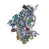
| Entry | Database: PDB / ID: 7v2o | ||||||||||||
|---|---|---|---|---|---|---|---|---|---|---|---|---|---|
| Title | T.thermophilus 30S ribosome with KsgA, class K4 | ||||||||||||
 Components Components |
| ||||||||||||
 Keywords Keywords | RIBOSOME / 30S subunit / KsgA / rRNA methyltransferase | ||||||||||||
| Function / homology |  Function and homology information Function and homology information16S rRNA (adenine1518-N6/adenine1519-N6)-dimethyltransferase / 16S rRNA (adenine(1518)-N(6)/adenine(1519)-N(6))-dimethyltransferase activity / rRNA (adenine-N6,N6-)-dimethyltransferase activity / rRNA methylation / ribosomal small subunit assembly / ribosomal small subunit biogenesis / small ribosomal subunit / small ribosomal subunit rRNA binding / cytosolic small ribosomal subunit / tRNA binding ...16S rRNA (adenine1518-N6/adenine1519-N6)-dimethyltransferase / 16S rRNA (adenine(1518)-N(6)/adenine(1519)-N(6))-dimethyltransferase activity / rRNA (adenine-N6,N6-)-dimethyltransferase activity / rRNA methylation / ribosomal small subunit assembly / ribosomal small subunit biogenesis / small ribosomal subunit / small ribosomal subunit rRNA binding / cytosolic small ribosomal subunit / tRNA binding / rRNA binding / structural constituent of ribosome / ribosome / translation / ribonucleoprotein complex / mRNA binding / RNA binding / zinc ion binding / metal ion binding / cytoplasm / cytosol Similarity search - Function | ||||||||||||
| Biological species |   Thermus thermophilus HB8 (bacteria) Thermus thermophilus HB8 (bacteria) | ||||||||||||
| Method | ELECTRON MICROSCOPY / single particle reconstruction / cryo EM / Resolution: 3.5 Å | ||||||||||||
 Authors Authors | Raina, R. / Singh, J. / Anand, R. / Vinothkumar, K.R. | ||||||||||||
| Funding support |  India, 3items India, 3items
| ||||||||||||
 Citation Citation | Journal: ACS Chem Biol / Year: 2022 Title: Decoding the Mechanism of Specific RNA Targeting by Ribosomal Methyltransferases. Authors: Juhi Singh / Rahul Raina / Kutti R Vinothkumar / Ruchi Anand / Abstract: Methylation of specific nucleotides is integral for ribosomal biogenesis and also serves as a common mechanism to confer antibiotic resistance by pathogenic bacteria. Here, by determining the high- ...Methylation of specific nucleotides is integral for ribosomal biogenesis and also serves as a common mechanism to confer antibiotic resistance by pathogenic bacteria. Here, by determining the high-resolution structure of the 30S-KsgA complex by cryo-electron microscopy, a state was captured, where KsgA juxtaposes between helices h44 and h45 of the 30S ribosome, separating them, thereby enabling remodeling of the surrounded rRNA and allowing the cognate site to enter the methylation pocket. With the structure as a guide, several mutant versions of the ribosomes, where interacting bases in the catalytic helix h45 and surrounding helices h44, h24, and h27, were mutated and evaluated for their methylation efficiency revealing factors that direct the enzyme to its cognate site with high fidelity. The biochemical studies show that the three-dimensional environment of the ribosome enables the interaction of select loop regions in KsgA with the ribosome helices paramount to maintain selectivity. | ||||||||||||
| History |
|
- Structure visualization
| Structure viewer | Molecule: MolmilJmol/JSmol |
|---|
- Downloads & links
-Download
| PDBx/mmCIF format | 7v2o.cif.gz | 1.3 MB | Display | PDBx/mmCIF format |
|---|---|---|---|---|
| PDB format | pdb7v2o.ent.gz | 1 MB | Display | PDB format |
| PDBx/mmJSON format | 7v2o.json.gz | Tree view | PDBx/mmJSON format | |
| Others |  Other downloads Other downloads |
-Validation report
| Arichive directory | https://data.pdbj.org/pub/pdb/validation_reports/v2/7v2oftp://data.pdbj.org/pub/pdb/validation_reports/v2/7v2o | HTTPS FTP |
|---|
-Related structure data
| Related structure data |  31658MC 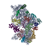 7v2lC 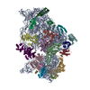 7v2mC 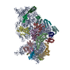 7v2nC 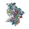 7v2pC 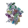 7v2qC C: citing same article ( M: map data used to model this data |
|---|---|
| Similar structure data |


-Links
 PDBj
PDBj


- Assembly
| Deposited unit | 
|
|---|---|
| 1 |
|
-Components
-30S ribosomal protein ... , 20 types, 20 molecules BCDEFGHIJKLMNOPQRSTV
| #2: Protein | Mass: 29317.703 Da / Num. of mol.: 1 / Source method: isolated from a natural source / Source: (natural) Thermus thermophilus HB8 (bacteria) / References: UniProt: P80371 |
|---|---|
| #3: Protein | Mass: 26751.076 Da / Num. of mol.: 1 / Source method: isolated from a natural source / Source: (natural) Thermus thermophilus HB8 (bacteria) / References: UniProt: P80372 |
| #4: Protein | Mass: 24373.447 Da / Num. of mol.: 1 / Source method: isolated from a natural source / Source: (natural) Thermus thermophilus HB8 (bacteria) / References: UniProt: P80373 |
| #5: Protein | Mass: 17583.416 Da / Num. of mol.: 1 / Source method: isolated from a natural source / Source: (natural) Thermus thermophilus HB8 (bacteria) / References: UniProt: Q5SHQ5 |
| #6: Protein | Mass: 11988.753 Da / Num. of mol.: 1 / Source method: isolated from a natural source / Source: (natural) Thermus thermophilus HB8 (bacteria) / References: UniProt: Q5SLP8 |
| #7: Protein | Mass: 18050.973 Da / Num. of mol.: 1 / Source method: isolated from a natural source / Source: (natural) Thermus thermophilus HB8 (bacteria) / References: UniProt: P17291 |
| #8: Protein | Mass: 15868.570 Da / Num. of mol.: 1 / Source method: isolated from a natural source / Source: (natural) Thermus thermophilus HB8 (bacteria) / References: UniProt: P0DOY9 |
| #9: Protein | Mass: 14410.614 Da / Num. of mol.: 1 / Source method: isolated from a natural source / Source: (natural) Thermus thermophilus HB8 (bacteria) / References: UniProt: P80374 |
| #10: Protein | Mass: 11954.968 Da / Num. of mol.: 1 / Source method: isolated from a natural source / Source: (natural) Thermus thermophilus HB8 (bacteria) / References: UniProt: Q5SHN7 |
| #11: Protein | Mass: 13737.868 Da / Num. of mol.: 1 / Source method: isolated from a natural source / Source: (natural) Thermus thermophilus HB8 (bacteria) / References: UniProt: P80376 |
| #12: Protein | Mass: 14637.384 Da / Num. of mol.: 1 / Source method: isolated from a natural source / Source: (natural) Thermus thermophilus HB8 (bacteria) / References: UniProt: Q5SHN3 |
| #13: Protein | Mass: 14338.861 Da / Num. of mol.: 1 / Source method: isolated from a natural source / Source: (natural) Thermus thermophilus HB8 (bacteria) / References: UniProt: P80377 |
| #14: Protein | Mass: 7158.725 Da / Num. of mol.: 1 / Source method: isolated from a natural source / Source: (natural) Thermus thermophilus HB8 (bacteria) / References: UniProt: P0DOY6 |
| #15: Protein | Mass: 10578.407 Da / Num. of mol.: 1 / Source method: isolated from a natural source / Source: (natural) Thermus thermophilus HB8 (bacteria) / References: UniProt: Q5SJ76 |
| #16: Protein | Mass: 10409.983 Da / Num. of mol.: 1 / Source method: isolated from a natural source / Source: (natural) Thermus thermophilus HB8 (bacteria) / References: UniProt: Q5SJH3 |
| #17: Protein | Mass: 12325.655 Da / Num. of mol.: 1 / Source method: isolated from a natural source / Source: (natural) Thermus thermophilus HB8 (bacteria) / References: UniProt: P0DOY7 |
| #18: Protein | Mass: 10258.299 Da / Num. of mol.: 1 / Source method: isolated from a natural source / Source: (natural) Thermus thermophilus HB8 (bacteria) / References: UniProt: Q5SLQ0 |
| #19: Protein | Mass: 10605.464 Da / Num. of mol.: 1 / Source method: isolated from a natural source / Source: (natural) Thermus thermophilus HB8 (bacteria) / References: UniProt: Q5SHP2 |
| #20: Protein | Mass: 11736.143 Da / Num. of mol.: 1 / Source method: isolated from a natural source / Source: (natural) Thermus thermophilus HB8 (bacteria) / References: UniProt: P80380 |
| #22: Protein/peptide | Mass: 3350.030 Da / Num. of mol.: 1 / Source method: isolated from a natural source / Source: (natural) Thermus thermophilus HB8 (bacteria) / References: UniProt: Q5SIH3 |
-RNA chain / Protein / Non-polymers , 3 types, 4 molecules AU
| #1: RNA chain | Mass: 493958.281 Da / Num. of mol.: 1 / Source method: isolated from a natural source / Source: (natural) Thermus thermophilus HB8 (bacteria) / References: GenBank: M26923.1 |
|---|---|
| #21: Protein | Mass: 33588.812 Da / Num. of mol.: 1 Source method: isolated from a genetically manipulated source Source: (gene. exp.) Gene: rsmA, ksgA, BSU00420 / Production host: References: UniProt: P37468, 16S rRNA (adenine1518-N6/adenine1519-N6)-dimethyltransferase |
| #23: Chemical |  Mass: 65.409 Da / Num. of mol.: 2 / Source method: obtained synthetically / Formula: Zn Mass: 65.409 Da / Num. of mol.: 2 / Source method: obtained synthetically / Formula: Zn |
-Details
| Has ligand of interest | N |
|---|
-Experimental details
-Experiment
| Experiment | Method: ELECTRON MICROSCOPY |
|---|---|
| EM experiment | Aggregation state: PARTICLE / 3D reconstruction method: single particle reconstruction |
- Sample preparation
| Component |
| ||||||||||||||||||||||||||||
|---|---|---|---|---|---|---|---|---|---|---|---|---|---|---|---|---|---|---|---|---|---|---|---|---|---|---|---|---|---|
| Molecular weight | Value: 0.74 MDa / Experimental value: NO | ||||||||||||||||||||||||||||
| Source (natural) |
| ||||||||||||||||||||||||||||
| Source (recombinant) | Organism: | ||||||||||||||||||||||||||||
| Buffer solution | pH: 7.5 | ||||||||||||||||||||||||||||
| Buffer component |
| ||||||||||||||||||||||||||||
| Specimen | Embedding applied: NO / Shadowing applied: NO / Staining applied: NO / Vitrification applied: YES Details: ~2 micromolar concentration of ribosome+KsgA was used during freezing | ||||||||||||||||||||||||||||
| Specimen support | Grid material: GOLD / Grid mesh size: 300 divisions/in. / Grid type: Quantifoil R1.2/1.3 | ||||||||||||||||||||||||||||
| Vitrification | Instrument: FEI VITROBOT MARK IV / Cryogen name: ETHANE / Humidity: 100 % / Chamber temperature: 277.15 K Details: After application of the complex, 15 seconds wait time was given before blotting. |
- Electron microscopy imaging
| Experimental equipment |  Model: Titan Krios / Image courtesy: FEI Company |
|---|---|
| Microscopy | Model: FEI TITAN KRIOS |
| Electron gun | Electron source:  FIELD EMISSION GUN / Accelerating voltage: 300 kV / Illumination mode: FLOOD BEAM FIELD EMISSION GUN / Accelerating voltage: 300 kV / Illumination mode: FLOOD BEAM |
| Electron lens | Mode: BRIGHT FIELD / Nominal magnification: 59000 X / Calibrated magnification: 101449 X / Nominal defocus max: 3500 nm / Nominal defocus min: 1800 nm / Cs: 2.7 mm / C2 aperture diameter: 50 µm / Alignment procedure: COMA FREE |
| Specimen holder | Cryogen: NITROGEN / Specimen holder model: FEI TITAN KRIOS AUTOGRID HOLDER |
| Image recording | Average exposure time: 2 sec. / Electron dose: 43 e/Å2 / Detector mode: INTEGRATING / Film or detector model: FEI FALCON III (4k x 4k) / Num. of grids imaged: 1 Details: Total number of frames is 30. Each frame had 1.43 e/A2 |
| Image scans | Sampling size: 14 µm / Width: 4096 / Height: 4096 |
- Processing
| EM software |
| |||||||||||||||||||||||||||||||||||||||||||||||||||||||
|---|---|---|---|---|---|---|---|---|---|---|---|---|---|---|---|---|---|---|---|---|---|---|---|---|---|---|---|---|---|---|---|---|---|---|---|---|---|---|---|---|---|---|---|---|---|---|---|---|---|---|---|---|---|---|---|---|
| CTF correction | Type: PHASE FLIPPING AND AMPLITUDE CORRECTION | |||||||||||||||||||||||||||||||||||||||||||||||||||||||
| Symmetry | Point symmetry: C1 (asymmetric) | |||||||||||||||||||||||||||||||||||||||||||||||||||||||
| 3D reconstruction | Resolution: 3.5 Å / Resolution method: FSC 0.143 CUT-OFF / Num. of particles: 64307 / Algorithm: FOURIER SPACE / Num. of class averages: 1 / Symmetry type: POINT | |||||||||||||||||||||||||||||||||||||||||||||||||||||||
| Atomic model building | B value: 109.3 / Protocol: OTHER / Space: RECIPROCAL Details: Phenix was used in real space for initial refinement. The final refinements was performed with REFMAC within ccpem | |||||||||||||||||||||||||||||||||||||||||||||||||||||||
| Atomic model building | PDB-ID: 4B3R Accession code: 4B3R / Source name: PDB / Type: experimental model |