 Movie
Movie Controller


+ Open data

- Basic information
| Entry | Database: PDB / ID: 7uwq | ||||||
|---|---|---|---|---|---|---|---|
| Title | Klebsiella pneumoniae adenosine monophosphate nucleosidase | ||||||
 Components Components | AMP nucleosidase | ||||||
 Keywords Keywords | HYDROLASE / nucleosidase / AMP / salvage | ||||||
| Function / homology |  Function and homology information Function and homology informationAMP nucleosidase / AMP nucleosidase activity / nucleoside metabolic process / AMP salvage / cytosol Similarity search - Function | ||||||
| Biological species |  Klebsiella pneumoniae (bacteria) Klebsiella pneumoniae (bacteria) | ||||||
| Method | ELECTRON MICROSCOPY / single particle reconstruction / cryo EM / Resolution: 3.05 Å | ||||||
 Authors Authors | Richardson, B.C. / French, J.B. | ||||||
| Funding support |  United States, 1items United States, 1items
| ||||||
 Citation Citation | Journal: PLoS One / Year: 2022 Title: Structure of Klebsiella pneumoniae adenosine monophosphate nucleosidase. Authors: Brian C Richardson / Roger Shek / Wesley C Van Voorhis / Jarrod B French / Abstract: Klebsiella pneumoniae is a bacterial pathogen that is increasingly responsible for hospital-acquired pneumonia and sepsis. Progressive development of antibiotic resistance has led to higher mortality ...Klebsiella pneumoniae is a bacterial pathogen that is increasingly responsible for hospital-acquired pneumonia and sepsis. Progressive development of antibiotic resistance has led to higher mortality rates and creates a need for novel treatments. Because of the essential role that nucleotides play in many bacterial processes, enzymes involved in purine and pyrimidine metabolism and transport are ideal targets for the development of novel antibiotics. Herein we describe the structure of K. pneumoniae adenosine monophosphate nucleosidase (KpAmn), a purine salvage enzyme unique to bacteria, as determined by cryoelectron microscopy. The data detail a well conserved fold with a hexameric overall structure and clear density for the putative active site residues. Comparison to the crystal structures of homologous prokaryotic proteins confirms the presence of many of the conserved structural features of this protein yet reveals differences in distal loops in the absence of crystal contacts. This first cryo-EM structure of an Amn enzyme provides a basis for future structure-guided drug development and extends the accuracy of structural characterization of this family of proteins beyond this clinically relevant organism. | ||||||
| History |
|
- Structure visualization
| Structure viewer | Molecule: MolmilJmol/JSmol |
|---|
- Downloads & links
-Download
| PDBx/mmCIF format | 7uwq.cif.gz | 457.5 KB | Display | PDBx/mmCIF format |
|---|---|---|---|---|
| PDB format | pdb7uwq.ent.gz | 368 KB | Display | PDB format |
| PDBx/mmJSON format | 7uwq.json.gz | Tree view | PDBx/mmJSON format | |
| Others |  Other downloads Other downloads |
-Validation report
| Arichive directory | https://data.pdbj.org/pub/pdb/validation_reports/uw/7uwqftp://data.pdbj.org/pub/pdb/validation_reports/uw/7uwq | HTTPS FTP |
|---|
-Related structure data
| Related structure data |  26838MC M: map data used to model this data C: citing same article ( |
|---|---|
| Similar structure data |
-Links
 PDBj
PDBj- Assembly
| Deposited unit | 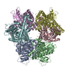
| ||||||||||||||||||||||||||||||||||||||||||||||||||||||||||||||||||||||||||||||||||||||||||||||
|---|---|---|---|---|---|---|---|---|---|---|---|---|---|---|---|---|---|---|---|---|---|---|---|---|---|---|---|---|---|---|---|---|---|---|---|---|---|---|---|---|---|---|---|---|---|---|---|---|---|---|---|---|---|---|---|---|---|---|---|---|---|---|---|---|---|---|---|---|---|---|---|---|---|---|---|---|---|---|---|---|---|---|---|---|---|---|---|---|---|---|---|---|---|---|---|
| 1 |
| ||||||||||||||||||||||||||||||||||||||||||||||||||||||||||||||||||||||||||||||||||||||||||||||
| Noncrystallographic symmetry (NCS) | NCS domain:
NCS domain segments:
NCS oper:
|
-Components
| #1: Protein | Mass: 55326.336 Da / Num. of mol.: 6 Source method: isolated from a genetically manipulated source Source: (gene. exp.) Klebsiella pneumoniae (bacteria)Gene: amn, A7B01_08185, B4U61_13745, B5L96_10595, BL124_00007990, BN49_3643, BS595_23510, C3F39_24645, DDJ63_13085, DRB11_00280, EAO17_04140, FXN67_19000, G5637_19470, G7Z27_09550, GJJ12_013280, ...Gene: amn, A7B01_08185, B4U61_13745, B5L96_10595, BL124_00007990, BN49_3643, BS595_23510, C3F39_24645, DDJ63_13085, DRB11_00280, EAO17_04140, FXN67_19000, G5637_19470, G7Z27_09550, GJJ12_013280, GNE24_03025, GNG14_09410, GPZ86_08955, HV479_00330, NCTC11679_01882, NCTC13443_04974, NCTC13465_03257, NCTC204_04420, NCTC9128_08085, NCTC9617_06427, NCTC9637_02601, SAMEA3499874_04631, SAMEA3499901_02408, SAMEA3500057_04230, SAMEA3512100_00855, SAMEA3538828_00203, SAMEA3649733_00677, SAMEA3649758_02509, SAMEA3720909_04803, SAMEA3727630_01134, SAMEA3727643_04047, SAMEA3727679_02343, SAMEA3729663_00178, SAMEA4364603_01617, SAMEA4873619_01962, SAMEA4873632_01182 Production host: |
|---|
-Experimental details
-Experiment
| Experiment | Method: ELECTRON MICROSCOPY |
|---|---|
| EM experiment | Aggregation state: PARTICLE / 3D reconstruction method: single particle reconstruction |
- Sample preparation
| Component | Name: Adenosine monophosphate nucleosidase / Type: COMPLEX / Entity ID: all / Source: RECOMBINANT | ||||||||||||||||||||||||||||||
|---|---|---|---|---|---|---|---|---|---|---|---|---|---|---|---|---|---|---|---|---|---|---|---|---|---|---|---|---|---|---|---|
| Molecular weight | Experimental value: NO | ||||||||||||||||||||||||||||||
| Source (natural) | Organism: Klebsiella pneumoniae (bacteria) | ||||||||||||||||||||||||||||||
| Source (recombinant) | Organism: | ||||||||||||||||||||||||||||||
| Buffer solution | pH: 7 | ||||||||||||||||||||||||||||||
| Buffer component |
| ||||||||||||||||||||||||||||||
| Specimen | Conc.: 2 mg/ml / Embedding applied: NO / Shadowing applied: NO / Staining applied: NO / Vitrification applied: YES | ||||||||||||||||||||||||||||||
| Specimen support | Grid material: GOLD / Grid type: C-flat-1.2/1.3 | ||||||||||||||||||||||||||||||
| Vitrification | Instrument: FEI VITROBOT MARK IV / Cryogen name: ETHANE / Humidity: 96 % / Chamber temperature: 298 K / Details: 9 second blot |
- Electron microscopy imaging
| Experimental equipment |  Model: Titan Krios / Image courtesy: FEI Company |
|---|---|
| Microscopy | Model: FEI TITAN KRIOS |
| Electron gun | Electron source:  FIELD EMISSION GUN / Accelerating voltage: 300 kV / Illumination mode: FLOOD BEAM FIELD EMISSION GUN / Accelerating voltage: 300 kV / Illumination mode: FLOOD BEAM |
| Electron lens | Mode: BRIGHT FIELD / Nominal magnification: 96000 X / Nominal defocus max: 2600 nm / Nominal defocus min: 1000 nm |
| Image recording | Electron dose: 60 e/Å2 / Film or detector model: FEI FALCON III (4k x 4k) / Num. of grids imaged: 1 / Num. of real images: 679 |
| Image scans | Width: 4096 / Height: 4096 |
- Processing
| Software |
| ||||||||||||||||||||||||||||||||||||
|---|---|---|---|---|---|---|---|---|---|---|---|---|---|---|---|---|---|---|---|---|---|---|---|---|---|---|---|---|---|---|---|---|---|---|---|---|---|
| EM software |
| ||||||||||||||||||||||||||||||||||||
| CTF correction | Type: PHASE FLIPPING AND AMPLITUDE CORRECTION | ||||||||||||||||||||||||||||||||||||
| Particle selection | Num. of particles selected: 1494578 | ||||||||||||||||||||||||||||||||||||
| Symmetry | Point symmetry: D3 (2x3 fold dihedral) | ||||||||||||||||||||||||||||||||||||
| 3D reconstruction | Resolution: 3.05 Å / Resolution method: FSC 0.143 CUT-OFF / Num. of particles: 114781 / Num. of class averages: 17 / Symmetry type: POINT | ||||||||||||||||||||||||||||||||||||
| Atomic model building | Protocol: FLEXIBLE FIT / Space: REAL | ||||||||||||||||||||||||||||||||||||
| Atomic model building | PDB-ID: 1T8R Accession code: 1T8R / Source name: PDB / Type: experimental model | ||||||||||||||||||||||||||||||||||||
| Refinement | Cross valid method: NONE Stereochemistry target values: GeoStd + Monomer Library + CDL v1.2 | ||||||||||||||||||||||||||||||||||||
| Displacement parameters | Biso mean: 82.81 Å2 | ||||||||||||||||||||||||||||||||||||
| Refine LS restraints |
| ||||||||||||||||||||||||||||||||||||
| Refine LS restraints NCS |
|

