+ データを開く
- 基本情報
基本情報
| 登録情報 | データベース: PDB / ID: 7utj | ||||||
|---|---|---|---|---|---|---|---|
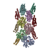
| タイトル | Cryogenic electron microscopy 3D map of F-actin bound by human dimeric alpha-catenin | ||||||
 要素 要素 |
| ||||||
 キーワード キーワード | CELL ADHESION / alpha-catenin / F-actin / F-actin binding protein / cell-cell junction | ||||||
| 機能・相同性 |  機能・相同性情報 機能・相同性情報negative regulation of integrin-mediated signaling pathway / CDH11 homotypic and heterotypic interactions / Regulation of CDH19 Expression and Function / Regulation of CDH11 function / gamma-catenin binding / epithelial cell-cell adhesion / zonula adherens / gap junction assembly / cellular response to indole-3-methanol / vinculin binding ...negative regulation of integrin-mediated signaling pathway / CDH11 homotypic and heterotypic interactions / Regulation of CDH19 Expression and Function / Regulation of CDH11 function / gamma-catenin binding / epithelial cell-cell adhesion / zonula adherens / gap junction assembly / cellular response to indole-3-methanol / vinculin binding / flotillin complex / negative regulation of cell motility / apical junction assembly / positive regulation of extrinsic apoptotic signaling pathway in absence of ligand / positive regulation of smoothened signaling pathway / Adherens junctions interactions / catenin complex / negative regulation of protein localization to nucleus / axon regeneration / cytoskeletal motor activator activity / negative regulation of neuroblast proliferation / smoothened signaling pathway / establishment or maintenance of cell polarity / Myogenesis / tropomyosin binding / mesenchyme migration / troponin I binding / myosin heavy chain binding / odontogenesis of dentin-containing tooth / filamentous actin / actin filament bundle / skeletal muscle thin filament assembly / striated muscle thin filament / actin filament bundle assembly / skeletal muscle myofibril / intercalated disc / actin monomer binding / negative regulation of extrinsic apoptotic signaling pathway in absence of ligand / neuroblast proliferation / RHO GTPases activate IQGAPs / skeletal muscle fiber development / stress fiber / ovarian follicle development / titin binding / extrinsic apoptotic signaling pathway in absence of ligand / actin filament polymerization / acrosomal vesicle / VEGFR2 mediated vascular permeability / filopodium / integrin-mediated signaling pathway / actin filament / adherens junction / 加水分解酵素; 酸無水物に作用; 酸無水物に作用・細胞または細胞小器官の運動に関与 / protein localization / beta-catenin binding / cell-cell adhesion / response to estrogen / calcium-dependent protein binding / male gonad development / cell-cell junction / actin filament binding / cell migration / actin cytoskeleton / lamellipodium / cell junction / cell body / hydrolase activity / cell adhesion / cadherin binding / protein domain specific binding / intracellular membrane-bounded organelle / focal adhesion / calcium ion binding / positive regulation of gene expression / structural molecule activity / magnesium ion binding / RNA binding / ATP binding / identical protein binding / plasma membrane / cytoplasm / cytosol 類似検索 - 分子機能 | ||||||
| 生物種 |   Homo sapiens (ヒト) Homo sapiens (ヒト) | ||||||
| 手法 | 電子顕微鏡法 / らせん対称体再構成法 / クライオ電子顕微鏡法 / 解像度: 2.77 Å | ||||||
 データ登録者 データ登録者 | Rangarajan, E.S. / Smith, E.W. / Izard, T. | ||||||
| 資金援助 |  米国, 1件 米国, 1件
| ||||||
 引用 引用 |  ジャーナル: Commun Biol / 年: 2023 ジャーナル: Commun Biol / 年: 2023タイトル: Distinct inter-domain interactions of dimeric versus monomeric α-catenin link cell junctions to filaments. 著者: Erumbi S Rangarajan / Emmanuel W Smith / Tina Izard /  要旨: Attachment between cells is crucial for almost all aspects of the life of cells. These inter-cell adhesions are mediated by the binding of transmembrane cadherin receptors of one cell to cadherins of ...Attachment between cells is crucial for almost all aspects of the life of cells. These inter-cell adhesions are mediated by the binding of transmembrane cadherin receptors of one cell to cadherins of a neighboring cell. Inside the cell, cadherin binds β-catenin, which interacts with α-catenin. The transitioning of cells between migration and adhesion is modulated by α-catenin, which links cell junctions and the plasma membrane to the actin cytoskeleton. At cell junctions, a single β-catenin/α-catenin heterodimer slips along filamentous actin in the direction of cytoskeletal tension which unfolds clustered heterodimers to form catch bonds with F-actin. Outside cell junctions, α-catenin dimerizes and links the plasma membrane to F-actin. Under cytoskeletal tension, α-catenin unfolds and forms an asymmetric catch bond with F-actin. To understand the mechanism of this important α-catenin function, we determined the 2.7 Å cryogenic electron microscopy (cryoEM) structures of filamentous actin alone and bound to human dimeric α-catenin. Our structures provide mechanistic insights into the role of the α-catenin interdomain interactions in directing α-catenin function and suggest a bivalent mechanism. Further, our cryoEM structure of human monomeric α-catenin provides mechanistic insights into α-catenin autoinhibition. Collectively, our structures capture the initial α-catenin interaction with F-actin before the sensing of force, which is a crucial event in cell adhesion and human disease. | ||||||
| 履歴 |
|
- 構造の表示
構造の表示
| 構造ビューア | 分子:  Molmil Molmil Jmol/JSmol Jmol/JSmol |
|---|
- ダウンロードとリンク
- ダウンロード
| PDBx/mmCIF形式 |  7utj.cif.gz 7utj.cif.gz | 602.5 KB | 表示 |  PDBx/mmCIF形式 PDBx/mmCIF形式 |
|---|---|---|---|---|
| PDB形式 |  pdb7utj.ent.gz pdb7utj.ent.gz | 459.2 KB | 表示 |  PDB形式 PDB形式 |
| PDBx/mmJSON形式 |  7utj.json.gz 7utj.json.gz | ツリー表示 |  PDBx/mmJSON形式 PDBx/mmJSON形式 | |
| その他 |  その他のダウンロード その他のダウンロード |
-検証レポート
| 文書・要旨 |  7utj_validation.pdf.gz 7utj_validation.pdf.gz | 1.6 MB | 表示 |  wwPDB検証レポート wwPDB検証レポート |
|---|---|---|---|---|
| 文書・詳細版 |  7utj_full_validation.pdf.gz 7utj_full_validation.pdf.gz | 1.6 MB | 表示 | |
| XML形式データ |  7utj_validation.xml.gz 7utj_validation.xml.gz | 94.7 KB | 表示 | |
| CIF形式データ |  7utj_validation.cif.gz 7utj_validation.cif.gz | 137.5 KB | 表示 | |
| アーカイブディレクトリ |  https://data.pdbj.org/pub/pdb/validation_reports/ut/7utj https://data.pdbj.org/pub/pdb/validation_reports/ut/7utj ftp://data.pdbj.org/pub/pdb/validation_reports/ut/7utj ftp://data.pdbj.org/pub/pdb/validation_reports/ut/7utj | HTTPS FTP |
-関連構造データ
| 関連構造データ |  26772MC  7uxfC M: このデータのモデリングに利用したマップデータ C: 同じ文献を引用 ( |
|---|---|
| 類似構造データ | 類似検索 - 機能・相同性  F&H 検索 F&H 検索 |
- リンク
リンク
- 集合体
集合体
| 登録構造単位 | 
| |||||||||||||||||||||||||||||||||||||||||||||||||||||||||||||||||||||||||||||||||||||||||||||||||||||||||||||||||||||||||||||||||||||||||||||
|---|---|---|---|---|---|---|---|---|---|---|---|---|---|---|---|---|---|---|---|---|---|---|---|---|---|---|---|---|---|---|---|---|---|---|---|---|---|---|---|---|---|---|---|---|---|---|---|---|---|---|---|---|---|---|---|---|---|---|---|---|---|---|---|---|---|---|---|---|---|---|---|---|---|---|---|---|---|---|---|---|---|---|---|---|---|---|---|---|---|---|---|---|---|---|---|---|---|---|---|---|---|---|---|---|---|---|---|---|---|---|---|---|---|---|---|---|---|---|---|---|---|---|---|---|---|---|---|---|---|---|---|---|---|---|---|---|---|---|---|---|---|---|
| 1 |
| |||||||||||||||||||||||||||||||||||||||||||||||||||||||||||||||||||||||||||||||||||||||||||||||||||||||||||||||||||||||||||||||||||||||||||||
| 非結晶学的対称性 (NCS) | NCSドメイン:
NCSドメイン領域:
|
 ムービー
ムービー コントローラー


 PDBj
PDBj


