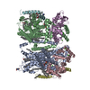
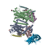
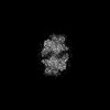
- PDB-7usw: Structure of Expanded C. elegans TMC-1 complex -
+
Open data
ID or keywords:
Loading...
-
Basic information
Entry
Database: PDB / ID: 7usw
Title
Structure of Expanded C. elegans TMC-1 complex
Components
(Transmembrane ...) x 2
CALMyrin (Calcium and Integrin Binding protein) homolog
Keywords
MEMBRANE PROTEIN / Complex
Function / homology
Function and homology information
detection of stimulus involved in sensory perception / striated muscle dense body / sensory perception of chemical stimulus / mechanosensitive monoatomic ion channel activity / non-motile cilium / sodium channel activity / monoatomic ion channel activity / calcium ion homeostasis / monoatomic ion transmembrane transport / neuronal cell body ...detection of stimulus involved in sensory perception / striated muscle dense body / sensory perception of chemical stimulus / mechanosensitive monoatomic ion channel activity / non-motile cilium / sodium channel activity / monoatomic ion channel activity / calcium ion homeostasis / monoatomic ion transmembrane transport / neuronal cell body / calcium ion binding / magnesium ion binding / plasma membrane / cytoplasm Similarity search - Function
Journal: Nature / Year: 2022 Title: Structures of the TMC-1 complex illuminate mechanosensory transduction. Authors: Hanbin Jeong / Sarah Clark / April Goehring / Sepehr Dehghani-Ghahnaviyeh / Ali Rasouli / Emad Tajkhorshid / Eric Gouaux / Abstract: The initial step in the sensory transduction pathway underpinning hearing and balance in mammals involves the conversion of force into the gating of a mechanosensory transduction channel. Despite the ...The initial step in the sensory transduction pathway underpinning hearing and balance in mammals involves the conversion of force into the gating of a mechanosensory transduction channel. Despite the profound socioeconomic impacts of hearing disorders and the fundamental biological significance of understanding mechanosensory transduction, the composition, structure and mechanism of the mechanosensory transduction complex have remained poorly characterized. Here we report the single-particle cryo-electron microscopy structure of the native transmembrane channel-like protein 1 (TMC-1) mechanosensory transduction complex isolated from Caenorhabditis elegans. The two-fold symmetric complex is composed of two copies each of the pore-forming TMC-1 subunit, the calcium-binding protein CALM-1 and the transmembrane inner ear protein TMIE. CALM-1 makes extensive contacts with the cytoplasmic face of the TMC-1 subunits, whereas the single-pass TMIE subunits reside on the periphery of the complex, poised like the handles of an accordion. A subset of complexes additionally includes a single arrestin-like protein, arrestin domain protein (ARRD-6), bound to a CALM-1 subunit. Single-particle reconstructions and molecular dynamics simulations show how the mechanosensory transduction complex deforms the membrane bilayer and suggest crucial roles for lipid-protein interactions in the mechanism by which mechanical force is transduced to ion channel gating.
History
Deposition
Apr 26, 2022
Deposition site: RCSB / Processing site: RCSB
Revision 1.0
Oct 19, 2022
Provider: repository / Type: Initial release
Revision 1.0
Oct 19, 2022
Data content type: EM metadata / Data content type: EM metadata / Provider: repository / Type: Initial release
Revision 1.0
Oct 19, 2022
Data content type: Half map / Part number: 1 / Data content type: Half map / Provider: repository / Type: Initial release
Revision 1.0
Oct 19, 2022
Data content type: Half map / Part number: 2 / Data content type: Half map / Provider: repository / Type: Initial release
Revision 1.0
Oct 19, 2022
Data content type: Image / Data content type: Image / Provider: repository / Type: Initial release
Revision 1.0
Oct 19, 2022
Data content type: Primary map / Data content type: Primary map / Provider: repository / Type: Initial release
Revision 1.0
Oct 19, 2022
Data content type: Half map / Part number: 1 / Data content type: Half map / Provider: repository / Type: Initial release
Revision 1.0
Oct 19, 2022
Data content type: Half map / Part number: 2 / Data content type: Half map / Provider: repository / Type: Initial release
Revision 1.0
Oct 19, 2022
Data content type: Image / Data content type: Image / Provider: repository / Type: Initial release
Revision 1.0
Oct 19, 2022
Data content type: Primary map / Data content type: Primary map / Provider: repository / Type: Initial release
Data content type: EM metadata / Data content type: EM metadata / EM metadata / Group: Data processing / Experimental summary / Data content type: EM metadata / EM metadata / Category: em_admin / em_software / Data content type: EM metadata / EM metadata / Item: _em_admin.last_update / _em_software.name
In the structure databanks used in Yorodumi, some data are registered as the other names, "COVID-19 virus" and "2019-nCoV". Here are the details of the virus and the list of structure data.
Jan 31, 2019. EMDB accession codes are about to change! (news from PDBe EMDB page)
EMDB accession codes are about to change! (news from PDBe EMDB page)
The allocation of 4 digits for EMDB accession codes will soon come to an end. Whilst these codes will remain in use, new EMDB accession codes will include an additional digit and will expand incrementally as the available range of codes is exhausted. The current 4-digit format prefixed with “EMD-” (i.e. EMD-XXXX) will advance to a 5-digit format (i.e. EMD-XXXXX), and so on. It is currently estimated that the 4-digit codes will be depleted around Spring 2019, at which point the 5-digit format will come into force.
The EM Navigator/Yorodumi systems omit the EMD- prefix.
Related info.:Q: What is EMD? / ID/Accession-code notation in Yorodumi/EM Navigator
Yorodumi is a browser for structure data from EMDB, PDB, SASBDB, etc.
This page is also the successor to EM Navigator detail page, and also detail information page/front-end page for Omokage search.
The word "yorodu" (or yorozu) is an old Japanese word meaning "ten thousand". "mi" (miru) is to see.
Related info.:EMDB / PDB / SASBDB / Comparison of 3 databanks / Yorodumi Search / Aug 31, 2016. New EM Navigator & Yorodumi / Yorodumi Papers / Jmol/JSmol / Function and homology information / Changes in new EM Navigator and Yorodumi
 Movie
Movie Controller


 Open data
 Basic information
 Components
 Keywords
 Function and homology information
 Authors
 United States, 1items
  Citation
 Structure visualization
 Downloads & links
 Other downloads
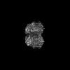

 PDBj
PDBj


 Assembly
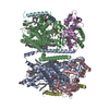

 Type: D-saccharide, beta linking / Mass: 221.208 Da / Num. of mol.: 2 / Source method: obtained synthetically / Formula: C8H15NO6 / Feature type: SUBJECT OF INVESTIGATION


 Mass: 40.078 Da / Num. of mol.: 8 / Source method: obtained synthetically / Formula: Ca / Feature type: SUBJECT OF INVESTIGATION
 Mass: 744.034 Da / Num. of mol.: 2 / Source method: obtained synthetically / Formula: C41H78NO8P / Feature type: SUBJECT OF INVESTIGATION / Comment: DOPE, phospholipid*YM
 Mass: 748.065 Da / Num. of mol.: 2 / Source method: obtained synthetically / Formula: C41H82NO8P / Feature type: SUBJECT OF INVESTIGATION / Comment: phospholipid*YM
 Mass: 172.308 Da / Num. of mol.: 34 / Source method: isolated from a natural source / Formula: C11H24O / Feature type: SUBJECT OF INVESTIGATION
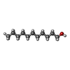
 Mass: 170.335 Da / Num. of mol.: 12 / Source method: isolated from a natural source / Formula: C12H26 / Feature type: SUBJECT OF INVESTIGATION
 Mass: 386.654 Da / Num. of mol.: 2 / Source method: obtained synthetically / Formula: C27H46O / Feature type: SUBJECT OF INVESTIGATION
 Mass: 226.441 Da / Num. of mol.: 4 / Source method: obtained synthetically / Formula: C16H34 / Feature type: SUBJECT OF INVESTIGATION
 Mass: 256.424 Da / Num. of mol.: 4 / Source method: obtained synthetically / Formula: C16H32O2 / Feature type: SUBJECT OF INVESTIGATION
 Sample preparation
 Electron microscopy imaging
 FIELD EMISSION GUN / Accelerating voltage: 300 kV / Illumination mode: FLOOD BEAM
 Processing