 Movie
Movie Controller

[English] 日本語
 Yorodumi
- PDB-7ujl: Bacteriophage Lambda Red-Beta N-terminal domain helical assembly ... -
+ Open data

- Basic information
| Entry | Database: PDB / ID: 7ujl | ||||||
|---|---|---|---|---|---|---|---|
| Title | Bacteriophage Lambda Red-Beta N-terminal domain helical assembly in complex with dsDNA | ||||||
 Components Components |
| ||||||
 Keywords Keywords | RECOMBINATION/DNA / Annealase / Synaptase / SSAP / Single-strand annealing protein / DNA annealing intermediate / Recombinase / Two-component recombinase / Viral / DNA-binding / RECOMBINATION-DNA complex | ||||||
| Function / homology | Bacteriophage lambda, Recombination protein bet / RecT family / RecT family / DNA recombination / DNA binding / DNA / DNA (> 10) / Recombination protein bet Function and homology information Function and homology information | ||||||
| Biological species |  Escherichia virus Lambda Escherichia virus Lambda | ||||||
| Method | ELECTRON MICROSCOPY / helical reconstruction / cryo EM / Resolution: 3.3 Å | ||||||
 Authors Authors | Newing, T.P. / Tolun, G. | ||||||
| Funding support |  Australia, 1items Australia, 1items
| ||||||
 Citation Citation | Journal: Nat Commun / Year: 2022 Title: Redβ annealase structure reveals details of oligomerization and λ Red-mediated homologous DNA recombination. Authors: Timothy P Newing / Jodi L Brewster / Lucy J Fitschen / James C Bouwer / Nikolas P Johnston / Haibo Yu / Gökhan Tolun / Abstract: The Redβ protein of the bacteriophage λ red recombination system is a model annealase which catalyzes single-strand annealing homologous DNA recombination. Here we present the structure of a ...The Redβ protein of the bacteriophage λ red recombination system is a model annealase which catalyzes single-strand annealing homologous DNA recombination. Here we present the structure of a helical oligomeric annealing intermediate of Redβ, consisting of N-terminal residues 1-177 bound to two complementary 27mer oligonucleotides, determined via cryogenic electron microscopy (cryo-EM) to a final resolution of 3.3 Å. The structure reveals a continuous binding groove which positions and stabilizes complementary DNA strands in a planar orientation to facilitate base pairing via a network of hydrogen bonding. Definition of the inter-subunit interface provides a structural basis for the propensity of Redβ to oligomerize into functionally significant long helical filaments, a trait shared by most annealases. Our cryo-EM structure and molecular dynamics simulations suggest that residues 133-138 form a flexible loop which modulates access to the binding groove. More than half a century after its discovery, this combination of structural and computational observations has allowed us to propose molecular mechanisms for the actions of the model annealase Redβ, a defining member of the Redβ/RecT protein family. | ||||||
| History |
|
- Structure visualization
| Structure viewer | Molecule: MolmilJmol/JSmol |
|---|
- Downloads & links
-Download
| PDBx/mmCIF format | 7ujl.cif.gz | 51.2 KB | Display | PDBx/mmCIF format |
|---|---|---|---|---|
| PDB format | pdb7ujl.ent.gz | 31.9 KB | Display | PDB format |
| PDBx/mmJSON format | 7ujl.json.gz | Tree view | PDBx/mmJSON format | |
| Others |  Other downloads Other downloads |
-Validation report
| Arichive directory | https://data.pdbj.org/pub/pdb/validation_reports/uj/7ujlftp://data.pdbj.org/pub/pdb/validation_reports/uj/7ujl | HTTPS FTP |
|---|
-Related structure data
| Related structure data | 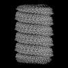 26566MC M: map data used to model this data C: citing same article ( |
|---|---|
| Similar structure data |
-Links
 PDBj
PDBj


- Assembly
| Deposited unit | 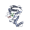
|
|---|---|
| 1 | x 60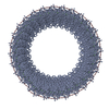
|
| 2 |
|
| Symmetry | Helical symmetry: (Circular symmetry: 1 / Dyad axis: no / N subunits divisor: 1 / Num. of operations: 60 / Rise per n subunits: 2.078 Å / Rotation per n subunits: -12.947 °) |
-Components
| #1: Protein | Mass: 21275.008 Da / Num. of mol.: 1 / Fragment: N-terminal domain (UNP residues 1-177) Source method: isolated from a genetically manipulated source Source: (gene. exp.) Escherichia virus Lambda / Gene: bet, betA, red-beta, redB / Plasmid: pET24aDetails (production host): pET24a containing the truncated N-terminal 177-residue fragment of the bet gene followed by a SSHHHHHH tag under the control of the lac repressor system Production host: |
|---|---|
| #2: DNA chain | Mass: 8244.295 Da / Num. of mol.: 1 / Source method: obtained synthetically / Source: (synth.) |
| #3: DNA chain | Mass: 8351.386 Da / Num. of mol.: 1 / Source method: obtained synthetically / Source: (synth.) |
| Has protein modification | N |
-Experimental details
-Experiment
| Experiment | Method: ELECTRON MICROSCOPY |
|---|---|
| EM experiment | Aggregation state: HELICAL ARRAY / 3D reconstruction method: helical reconstruction |
- Sample preparation
| Component |
| |||||||||||||||||||||||||||||||||||
|---|---|---|---|---|---|---|---|---|---|---|---|---|---|---|---|---|---|---|---|---|---|---|---|---|---|---|---|---|---|---|---|---|---|---|---|---|
| Molecular weight |
| |||||||||||||||||||||||||||||||||||
| Source (natural) |
| |||||||||||||||||||||||||||||||||||
| Source (recombinant) |
| |||||||||||||||||||||||||||||||||||
| Buffer solution | pH: 6 | |||||||||||||||||||||||||||||||||||
| Buffer component |
| |||||||||||||||||||||||||||||||||||
| Specimen | Conc.: 2.6 mg/ml / Embedding applied: NO / Shadowing applied: NO / Staining applied: NO / Vitrification applied: YES | |||||||||||||||||||||||||||||||||||
| Specimen support | Grid material: GOLD / Grid mesh size: 300 divisions/in. / Grid type: Quantifoil R1.2/1.3 | |||||||||||||||||||||||||||||||||||
| Vitrification | Instrument: FEI VITROBOT MARK IV / Cryogen name: ETHANE / Humidity: 100 % / Chamber temperature: 298 K Details: Sample loading volume ranged between 2 and 3 microlitres. Samples were blotted for 5 seconds prior to vitrification. |
- Electron microscopy imaging
| Experimental equipment |  Model: Titan Krios / Image courtesy: FEI Company |
|---|---|
| Microscopy | Model: FEI TITAN KRIOS |
| Electron gun | Electron source:  FIELD EMISSION GUN / Accelerating voltage: 300 kV / Illumination mode: FLOOD BEAM FIELD EMISSION GUN / Accelerating voltage: 300 kV / Illumination mode: FLOOD BEAM |
| Electron lens | Mode: BRIGHT FIELD / Calibrated magnification: 59500 X / Nominal defocus max: 2500 nm / Nominal defocus min: 500 nm / Cs: 2.7 mm |
| Specimen holder | Cryogen: NITROGEN / Specimen holder model: FEI TITAN KRIOS AUTOGRID HOLDER |
| Image recording | Average exposure time: 9 sec. / Electron dose: 50 e/Å2 / Detector mode: COUNTING / Film or detector model: GATAN K2 SUMMIT (4k x 4k) / Num. of grids imaged: 1 / Num. of real images: 4710 |
| EM imaging optics | Energyfilter name: GIF Quantum LS / Details: Installed but not used |
| Image scans | Width: 3838 / Height: 3710 / Movie frames/image: 50 / Used frames/image: 1-50 |
- Processing
| Software | Name: UCSF ChimeraX / Version: 1.3/v9 / Classification: model building / URL: https://www.rbvi.ucsf.edu/chimerax/ / Os: Windows / Type: package | ||||||||||||||||||||||||||||||||||||||||
|---|---|---|---|---|---|---|---|---|---|---|---|---|---|---|---|---|---|---|---|---|---|---|---|---|---|---|---|---|---|---|---|---|---|---|---|---|---|---|---|---|---|
| EM software |
| ||||||||||||||||||||||||||||||||||||||||
| CTF correction | Type: PHASE FLIPPING AND AMPLITUDE CORRECTION | ||||||||||||||||||||||||||||||||||||||||
| Helical symmerty | Angular rotation/subunit: -12.947 ° / Axial rise/subunit: 2.078 Å / Axial symmetry: C1 | ||||||||||||||||||||||||||||||||||||||||
| Particle selection | Num. of particles selected: 922280 Details: 922280 particles were initially selected using cryoSPARC filament tracing | ||||||||||||||||||||||||||||||||||||||||
| 3D reconstruction | Resolution: 3.3 Å / Resolution method: FSC 0.143 CUT-OFF / Num. of particles: 26525 / Algorithm: FOURIER SPACE / Num. of class averages: 2 / Symmetry type: HELICAL | ||||||||||||||||||||||||||||||||||||||||
| Atomic model building | Protocol: FLEXIBLE FIT / Space: REAL / Target criteria: Correlation coefficient |
