 ムービー
ムービー コントローラー


+ データを開く

- 基本情報
基本情報
| 登録情報 | データベース: PDB / ID: 7ubb | ||||||
|---|---|---|---|---|---|---|---|
| タイトル | Structure of RecT protein from Listeria innoccua phage A118 in complex with 83-mer ssDNA | ||||||
 要素 要素 | RecT | ||||||
 キーワード キーワード | DNA BINDING PROTEIN / DNA Recombination / DNA Annealing | ||||||
| 機能・相同性 | DNA single-strand annealing protein RecT / RecT family / RecT family / DNA metabolic process / DNA binding / Recombinase [Bacteriophage A118] 機能・相同性情報 機能・相同性情報 | ||||||
| 生物種 |  Listeria innocua Clip11262 (バクテリア) Listeria innocua Clip11262 (バクテリア) | ||||||
| 手法 | 電子顕微鏡法 / 単粒子再構成法 / クライオ電子顕微鏡法 / 解像度: 4.5 Å | ||||||
 データ登録者 データ登録者 | Bell, C.E. / Caldwell, B.J. | ||||||
| 資金援助 |  米国, 1件 米国, 1件
| ||||||
 引用 引用 | ジャーナル: Nat Commun / 年: 2022 タイトル: Structure of a RecT/Redβ family recombinase in complex with a duplex intermediate of DNA annealing. 著者: Brian J Caldwell / Andrew S Norris / Caroline F Karbowski / Alyssa M Wiegand / Vicki H Wysocki / Charles E Bell / 要旨: Some bacteriophage encode a recombinase that catalyzes single-stranded DNA annealing (SSA). These proteins are apparently related to RAD52, the primary human SSA protein. The best studied protein, ...Some bacteriophage encode a recombinase that catalyzes single-stranded DNA annealing (SSA). These proteins are apparently related to RAD52, the primary human SSA protein. The best studied protein, Redβ from bacteriophage λ, binds weakly to ssDNA, not at all to dsDNA, but tightly to a duplex intermediate of annealing formed when two complementary DNA strands are added to the protein sequentially. We used single particle cryo-electron microscopy (cryo-EM) to determine a 3.4 Å structure of a Redβ homolog from a prophage of Listeria innocua in complex with two complementary 83mer oligonucleotides. The structure reveals a helical protein filament bound to a DNA duplex that is highly extended and unwound. Native mass spectrometry confirms that the complex seen by cryo-EM is the predominant species in solution. The protein shares a common core fold with RAD52 and a similar mode of ssDNA-binding. These data provide insights into the mechanism of protein-catalyzed SSA. | ||||||
| 履歴 |
|
- 構造の表示
構造の表示
| 構造ビューア | 分子: MolmilJmol/JSmol |
|---|
- ダウンロードとリンク
-ダウンロード
| PDBx/mmCIF形式 | 7ubb.cif.gz | 186.7 KB | 表示 | PDBx/mmCIF形式 |
|---|---|---|---|---|
| PDB形式 | pdb7ubb.ent.gz | 141.8 KB | 表示 | PDB形式 |
| PDBx/mmJSON形式 | 7ubb.json.gz | ツリー表示 | PDBx/mmJSON形式 | |
| その他 |  その他のダウンロード その他のダウンロード |
-検証レポート
| 文書・要旨 | 7ubb_validation.pdf.gz | 1.5 MB | 表示 | wwPDB検証レポート |
|---|---|---|---|---|
| 文書・詳細版 | 7ubb_full_validation.pdf.gz | 1.5 MB | 表示 | |
| XML形式データ | 7ubb_validation.xml.gz | 36.7 KB | 表示 | |
| CIF形式データ | 7ubb_validation.cif.gz | 55.4 KB | 表示 | |
| アーカイブディレクトリ | https://data.pdbj.org/pub/pdb/validation_reports/ub/7ubbftp://data.pdbj.org/pub/pdb/validation_reports/ub/7ubb | HTTPS FTP |
-関連構造データ


-リンク
 PDBj
PDBj- 集合体
集合体
| 登録構造単位 | 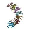
|
|---|---|
| 1 |
|
-要素
| #1: タンパク質 | 分子量: 30939.100 Da / 分子数: 8 / 由来タイプ: 組換発現 詳細: The modeled complex consists of 8 subunits of the RecT protein, which form a helical filament on ssDNA. The ssDNA was not modeled due to the low resolution. Density for four additional ...詳細: The modeled complex consists of 8 subunits of the RecT protein, which form a helical filament on ssDNA. The ssDNA was not modeled due to the low resolution. Density for four additional subunits, two on either end of the filament, was present, but not clear enough to dock into. 由来: (組換発現) Listeria innocua Clip11262 (バクテリア)株: ATCC BAA-680 / CLIP 11262 / 遺伝子: lin0085 / 発現宿主: 構成要素の詳細 | The modeled complex consists of 8 subunits of the RecT protein, which form a helical filament on ...The modeled complex consists of 8 subunits of the RecT protein, which form a helical filament on ssDNA. The ssDNA was not modeled due to the low resolution. Density for four additional subunits, two on either end of the filament, was present, but not clear enough to dock into. | |
|---|
-実験情報
-実験
| 実験 | 手法: 電子顕微鏡法 |
|---|---|
| EM実験 | 試料の集合状態: PARTICLE / 3次元再構成法: 単粒子再構成法 |
- 試料調製
試料調製
| 構成要素 | 名称: RecT protein from Listeria innocua phage A118, complexed with and 83-mer ssDNA タイプ: COMPLEX 詳細: The protein was purified by Nickel affinity and anion exchange chromatography. The DNA was chemically synthesized and HPLC purified. Entity ID: all / 由来: RECOMBINANT | ||||||||||||||||||||
|---|---|---|---|---|---|---|---|---|---|---|---|---|---|---|---|---|---|---|---|---|---|
| 分子量 | 値: 0.602 MDa / 実験値: NO | ||||||||||||||||||||
| 由来(天然) | 生物種: Listeria innocua Clip11262 (バクテリア) | ||||||||||||||||||||
| 由来(組換発現) | 生物種: | ||||||||||||||||||||
| 緩衝液 | pH: 6 詳細: The LiRecT protein was mixed at 37C with one oligonucleotide, and placed on ice for 90 min. Then immediately prior to vitrification, 1 ul of 1.5 mM n-dodecyl-beta-maltoside (Anatrace) was added (0.5 CMC). | ||||||||||||||||||||
| 緩衝液成分 |
| ||||||||||||||||||||
| 試料 | 濃度: 0.7 mg/ml / 包埋: NO / シャドウイング: NO / 染色: NO / 凍結: YES / 詳細: This sample was monodisperse | ||||||||||||||||||||
| 試料支持 | 詳細: 60 seconds at 20 mA using a Pelco easiGlow / グリッドの材料: GOLD / グリッドのサイズ: 300 divisions/in. / グリッドのタイプ: Quantifoil R1.2/1.3 | ||||||||||||||||||||
| 急速凍結 | 装置: FEI VITROBOT MARK IV / 凍結剤: ETHANE / 湿度: 100 % / 凍結前の試料温度: 277 K / 詳細: 1.5 second blot time. Ted Pella 595 filter paper. |
- 電子顕微鏡撮影
| 実験機器 |  モデル: Titan Krios / 画像提供: FEI Company |
|---|---|
| 顕微鏡 | モデル: FEI TITAN KRIOS |
| 電子銃 | 電子線源:  FIELD EMISSION GUN / 加速電圧: 300 kV / 照射モード: SPOT SCAN FIELD EMISSION GUN / 加速電圧: 300 kV / 照射モード: SPOT SCAN |
| 電子レンズ | モード: BRIGHT FIELD / 倍率(公称値): 81000 X / 最大 デフォーカス(公称値): 1000 nm / 最小 デフォーカス(公称値): 3500 nm / C2レンズ絞り径: 50 µm |
| 撮影 | 平均露光時間: 2.83 sec. / 電子線照射量: 65 e/Å2 フィルム・検出器のモデル: GATAN K3 BIOQUANTUM (6k x 4k) 撮影したグリッド数: 1 / 実像数: 1619 / 詳細: 45 fractions, 22.80 e-/A2/s |
| 電子光学装置 | エネルギーフィルター名称: GIF Bioquantum / 詳細: Gatan BioContinuum energy filter / エネルギーフィルタースリット幅: 20 eV / 球面収差補正装置: Cs corrector was used |
| 画像スキャン | 横: 6000 / 縦: 4000 |
- 解析
解析
| ソフトウェア | 名称: PHENIX / バージョン: 1.20.1_4487: / 分類: 精密化 | ||||||||||||||||||||||||
|---|---|---|---|---|---|---|---|---|---|---|---|---|---|---|---|---|---|---|---|---|---|---|---|---|---|
| EMソフトウェア |
| ||||||||||||||||||||||||
| CTF補正 | 詳細: Patch CTD estimation was implemented in cryoSPARC with am amplitude contrast of 0.1 タイプ: PHASE FLIPPING AND AMPLITUDE CORRECTION | ||||||||||||||||||||||||
| 粒子像の選択 | 選択した粒子像数: 1000 | ||||||||||||||||||||||||
| 対称性 | 点対称性: C1 (非対称) | ||||||||||||||||||||||||
| 3次元再構成 | 解像度: 4.5 Å / 解像度の算出法: FSC 0.143 CUT-OFF / 粒子像の数: 180965 詳細: The final resolution with a tight mask was 4.5 Angstrom 対称性のタイプ: POINT | ||||||||||||||||||||||||
| 原子モデル構築 | プロトコル: AB INITIO MODEL / 空間: REAL 詳細: Phenix Real Space Refinement was by rigid body refinement of the eight different subunits. Due to the limited resolution, the refinement was limited to rigid body only. |
