ムービー
ムービー コントローラー


+ データを開く

- 基本情報
基本情報
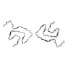
| 登録情報 | データベース: PDB / ID: 7uak | |||||||||
|---|---|---|---|---|---|---|---|---|---|---|
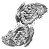
| タイトル | Structure of recombinantly assembled A53E alpha-synuclein fibrils | |||||||||
 要素 要素 | Alpha-synuclein | |||||||||
 キーワード キーワード | PROTEIN FIBRIL / alpha-synuclein fibrils / A53E mutant / cryo-EM structure / structural protein | |||||||||
| 機能・相同性 |  機能・相同性情報 機能・相同性情報negative regulation of mitochondrial electron transport, NADH to ubiquinone / neutral lipid metabolic process / regulation of phospholipase activity / negative regulation of monooxygenase activity / regulation of acyl-CoA biosynthetic process / negative regulation of dopamine uptake involved in synaptic transmission / negative regulation of norepinephrine uptake / positive regulation of glutathione peroxidase activity / positive regulation of SNARE complex assembly / positive regulation of hydrogen peroxide catabolic process ...negative regulation of mitochondrial electron transport, NADH to ubiquinone / neutral lipid metabolic process / regulation of phospholipase activity / negative regulation of monooxygenase activity / regulation of acyl-CoA biosynthetic process / negative regulation of dopamine uptake involved in synaptic transmission / negative regulation of norepinephrine uptake / positive regulation of glutathione peroxidase activity / positive regulation of SNARE complex assembly / positive regulation of hydrogen peroxide catabolic process / supramolecular fiber / negative regulation of transporter activity / mitochondrial membrane organization / negative regulation of chaperone-mediated autophagy / regulation of reactive oxygen species biosynthetic process / positive regulation of protein localization to cell periphery / regulation of synaptic vesicle recycling / negative regulation of platelet-derived growth factor receptor signaling pathway / negative regulation of exocytosis / regulation of glutamate secretion / regulation of norepinephrine uptake / response to iron(II) ion / SNARE complex assembly / positive regulation of neurotransmitter secretion / dopamine biosynthetic process / regulation of locomotion / positive regulation of inositol phosphate biosynthetic process / synaptic vesicle priming / regulation of macrophage activation / negative regulation of microtubule polymerization / dopamine uptake involved in synaptic transmission / synaptic vesicle transport / dynein complex binding / positive regulation of receptor recycling / regulation of dopamine secretion / protein kinase inhibitor activity / negative regulation of thrombin-activated receptor signaling pathway / response to type II interferon / cuprous ion binding / synaptic vesicle exocytosis / positive regulation of exocytosis / kinesin binding / positive regulation of endocytosis / mitochondrial ATP synthesis coupled electron transport / cysteine-type endopeptidase inhibitor activity involved in apoptotic process / response to magnesium ion / regulation of presynapse assembly / synaptic vesicle endocytosis / negative regulation of serotonin uptake / alpha-tubulin binding / localization / phospholipid metabolic process / supramolecular fiber organization / axon terminus / inclusion body / cellular response to copper ion / cellular response to epinephrine stimulus / Hsp70 protein binding / excitatory postsynaptic potential / response to interleukin-1 / adult locomotory behavior / SNARE binding / positive regulation of release of sequestered calcium ion into cytosol / fatty acid metabolic process / long-term synaptic potentiation / phosphoprotein binding / protein tetramerization / regulation of transmembrane transporter activity / synapse organization / regulation of long-term neuronal synaptic plasticity / microglial cell activation / negative regulation of protein kinase activity / protein destabilization / negative regulation of cysteine-type endopeptidase activity involved in apoptotic process / ferrous iron binding / tau protein binding / PKR-mediated signaling / positive regulation of protein serine/threonine kinase activity / receptor internalization / phospholipid binding / synaptic vesicle membrane / positive regulation of inflammatory response / activation of cysteine-type endopeptidase activity involved in apoptotic process / actin cytoskeleton / positive regulation of peptidyl-serine phosphorylation / actin binding / cell cortex / histone binding / cellular response to oxidative stress / growth cone / postsynapse / chemical synaptic transmission / neuron apoptotic process / negative regulation of neuron apoptotic process / amyloid fibril formation / response to lipopolysaccharide / molecular adaptor activity / oxidoreductase activity / lysosome / transcription cis-regulatory region binding 類似検索 - 分子機能 | |||||||||
| 生物種 |  Homo sapiens (ヒト) Homo sapiens (ヒト) | |||||||||
| 手法 | 電子顕微鏡法 / らせん対称体再構成法 / クライオ電子顕微鏡法 / 解像度: 3.38 Å | |||||||||
 データ登録者 データ登録者 | Zhou, K. / Zhou, H. | |||||||||
| 資金援助 |  米国, 2件 米国, 2件
| |||||||||
 引用 引用 | ジャーナル: J Biol Chem / 年: 2023 タイトル: Cryo-EM structure of amyloid fibril formed by α-synuclein hereditary A53E mutation reveals a distinct protofilament interface. 著者: Chuanqi Sun / Kang Zhou / Peter DePaola / Woo Shik Shin / Trae Hillyer / Michael R Sawaya / Ruowei Zhu / Chao Peng / Z Hong Zhou / Lin Jiang / 要旨: Synucleinopathies like Parkinson's disease (PD), dementia with Lewy bodies (DLB), and multiple systems atrophy (MSA), have the same pathologic feature of misfolded α-synuclein protein (α-syn) ...Synucleinopathies like Parkinson's disease (PD), dementia with Lewy bodies (DLB), and multiple systems atrophy (MSA), have the same pathologic feature of misfolded α-synuclein protein (α-syn) accumulation in the brain. PD patients who carry α-syn hereditary mutations tend to have earlier onset and more severe clinical symptoms than sporadic PD patients. Therefore, revealing the effect of hereditary mutations to the α-syn fibril structure can help us understand these synucleinopathies' structural basis. Here, we present a 3.38 Å cryo-electron microscopy structure of α-synuclein fibrils containing the hereditary A53E mutation. The A53E fibril is symmetrically composed of two protofilaments, similar to other fibril structures of WT and mutant α-synuclein. The new structure is distinct from all other synuclein fibrils, not only at the interface between proto-filaments, but also between residues packed within the same proto-filament. A53E has the smallest interface with the least buried surface area among all α-syn fibrils, consisting of only two contacting residues. Within the same protofilament, A53E reveals distinct residue re-arrangement and structural variation at a cavity near its fibril core. Moreover, the A53E fibrils exhibit slower fibril formation and lower stability compared to WT and other mutants like A53T and H50Q, while also demonstrate strong cellular seeding in α-synuclein biosensor cells and primary neurons. In summary, our study aims to highlight structural differences - both within and between the protofilaments of A53E fibrils - and interpret fibril formation and cellular seeding of α-synuclein pathology in disease, which could further our understanding of the structure-activity relationship of α-synuclein mutants. | |||||||||
| 履歴 |
|
- 構造の表示
構造の表示
| 構造ビューア | 分子: MolmilJmol/JSmol |
|---|
- ダウンロードとリンク
-ダウンロード
| PDBx/mmCIF形式 | 7uak.cif.gz | 72.1 KB | 表示 | PDBx/mmCIF形式 |
|---|---|---|---|---|
| PDB形式 | pdb7uak.ent.gz | 51.8 KB | 表示 | PDB形式 |
| PDBx/mmJSON形式 | 7uak.json.gz | ツリー表示 | PDBx/mmJSON形式 | |
| その他 |  その他のダウンロード その他のダウンロード |
-検証レポート
| 文書・要旨 | 7uak_validation.pdf.gz | 981.7 KB | 表示 | wwPDB検証レポート |
|---|---|---|---|---|
| 文書・詳細版 | 7uak_full_validation.pdf.gz | 987.9 KB | 表示 | |
| XML形式データ | 7uak_validation.xml.gz | 22.8 KB | 表示 | |
| CIF形式データ | 7uak_validation.cif.gz | 31.1 KB | 表示 | |
| アーカイブディレクトリ | https://data.pdbj.org/pub/pdb/validation_reports/ua/7uakftp://data.pdbj.org/pub/pdb/validation_reports/ua/7uak | HTTPS FTP |
-関連構造データ
-リンク
 PDBj
PDBj- 集合体
集合体
| 登録構造単位 | 
|
|---|---|
| 1 |
|
-要素
| #1: タンパク質 | 分子量: 14534.146 Da / 分子数: 6 / 変異: A53E / 由来タイプ: 組換発現 / 由来: (組換発現) Homo sapiens (ヒト) / 遺伝子: SNCA, NACP, PARK1発現宿主:  参照: UniProt: P37840 |
|---|
-実験情報
-実験
| 実験 | 手法: 電子顕微鏡法 |
|---|---|
| EM実験 | 試料の集合状態: FILAMENT / 3次元再構成法: らせん対称体再構成法 |
- 試料調製
試料調製
| 構成要素 | 名称: alpha-synuclein A53E mutant / タイプ: ORGANELLE OR CELLULAR COMPONENT / Entity ID: all / 由来: RECOMBINANT |
|---|---|
| 分子量 | 値: 59 kDa/nm / 実験値: NO |
| 由来(天然) | 生物種: Homo sapiens (ヒト) |
| 由来(組換発現) | 生物種: |
| 緩衝液 | pH: 7.4 |
| 緩衝液成分 | 濃度: 15 mM / 名称: tetrabutylphosphonium bromide / 式: TBPB |
| 試料 | 濃度: 5 mg/ml / 包埋: NO / シャドウイング: NO / 染色: NO / 凍結: YES |
| 試料支持 | グリッドの材料: COPPER / グリッドのサイズ: 300 divisions/in. / グリッドのタイプ: Quantifoil R2/1 |
| 急速凍結 | 装置: FEI VITROBOT MARK IV / 凍結剤: ETHANE / 湿度: 100 % / 凍結前の試料温度: 283.15 K |
- 電子顕微鏡撮影
| 実験機器 |  モデル: Titan Krios / 画像提供: FEI Company |
|---|---|
| 顕微鏡 | モデル: FEI TITAN KRIOS |
| 電子銃 | 電子線源:  FIELD EMISSION GUN / 加速電圧: 300 kV / 照射モード: FLOOD BEAM FIELD EMISSION GUN / 加速電圧: 300 kV / 照射モード: FLOOD BEAM |
| 電子レンズ | モード: BRIGHT FIELD / 倍率(公称値): 130000 X / 最大 デフォーカス(公称値): 3000 nm / 最小 デフォーカス(公称値): 1500 nm / Calibrated defocus min: 1500 nm / 最大 デフォーカス(補正後): 3000 nm / Cs: 2.7 mm / C2レンズ絞り径: 70 µm / アライメント法: COMA FREE |
| 試料ホルダ | 凍結剤: NITROGEN 試料ホルダーモデル: FEI TITAN KRIOS AUTOGRID HOLDER |
| 撮影 | 平均露光時間: 7 sec. / 電子線照射量: 48 e/Å2 / 検出モード: SUPER-RESOLUTION フィルム・検出器のモデル: GATAN K2 SUMMIT (4k x 4k) 撮影したグリッド数: 1 / 実像数: 2767 |
| 電子光学装置 | エネルギーフィルター名称: GIF Quantum LS / エネルギーフィルタースリット幅: 20 eV |
| 画像スキャン | 横: 3838 / 縦: 3710 / 動画フレーム数/画像: 5 / 利用したフレーム数/画像: 2-34 |
- 解析
解析
| EMソフトウェア |
| ||||||||||||||||||||||||||||
|---|---|---|---|---|---|---|---|---|---|---|---|---|---|---|---|---|---|---|---|---|---|---|---|---|---|---|---|---|---|
| CTF補正 | タイプ: PHASE FLIPPING AND AMPLITUDE CORRECTION | ||||||||||||||||||||||||||||
| らせん対称 | 回転角度/サブユニット: 179.504 ° / 軸方向距離/サブユニット: 2.418 Å / らせん対称軸の対称性: C1 | ||||||||||||||||||||||||||||
| 粒子像の選択 | 選択した粒子像数: 46626 | ||||||||||||||||||||||||||||
| 3次元再構成 | 解像度: 3.38 Å / 解像度の算出法: FSC 0.143 CUT-OFF / 粒子像の数: 31916 / アルゴリズム: SIMULTANEOUS ITERATIVE (SIRT) / クラス平均像の数: 1 / 対称性のタイプ: HELICAL | ||||||||||||||||||||||||||||
| 原子モデル構築 | B value: 125 / プロトコル: RIGID BODY FIT / 空間: REAL | ||||||||||||||||||||||||||||
| 原子モデル構築 | PDB-ID: 6LRQ PDB chain-ID: A / Accession code: 6LRQ / Source name: PDB / タイプ: experimental model |