 Movie
Movie Controller

[English] 日本語
 Yorodumi
- PDB-7u12: TMEM106B(120-254) singlet amyloid fibril from frontotemporal loba... -
+ Open data

- Basic information
| Entry | Database: PDB / ID: 7u12 | |||||||||||||||||||||||||||
|---|---|---|---|---|---|---|---|---|---|---|---|---|---|---|---|---|---|---|---|---|---|---|---|---|---|---|---|---|
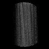
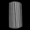
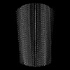
| Title | TMEM106B(120-254) singlet amyloid fibril from frontotemporal lobar degeneration with TDP-43 pathology (FTLD-TDP) type A (case 2) | |||||||||||||||||||||||||||
 Components Components | Transmembrane protein 106B | |||||||||||||||||||||||||||
 Keywords Keywords | PROTEIN FIBRIL / TMEM106B / Amyloid fibril | |||||||||||||||||||||||||||
| Function / homology |  Function and homology information Function and homology informationlysosomal protein catabolic process / lysosomal lumen acidification / lysosome localization / regulation of lysosome organization / positive regulation of dendrite development / lysosomal transport / dendrite morphogenesis / lysosome organization / neuron cellular homeostasis / late endosome membrane ...lysosomal protein catabolic process / lysosomal lumen acidification / lysosome localization / regulation of lysosome organization / positive regulation of dendrite development / lysosomal transport / dendrite morphogenesis / lysosome organization / neuron cellular homeostasis / late endosome membrane / ATPase binding / lysosome / endosome / lysosomal membrane / plasma membrane Similarity search - Function | |||||||||||||||||||||||||||
| Biological species |  Homo sapiens (human) Homo sapiens (human) | |||||||||||||||||||||||||||
| Method | ELECTRON MICROSCOPY / helical reconstruction / cryo EM / Resolution: 3.5 Å | |||||||||||||||||||||||||||
 Authors Authors | Fitzpatrick, A.W.P. / Stowell, M.H.B. / Chang, A. / Xiang, X. / Wang, J. / Lee, C. / Arakhamia, T. / Simjanoska, M. / Wang, C. / Carlomagno, Y. ...Fitzpatrick, A.W.P. / Stowell, M.H.B. / Chang, A. / Xiang, X. / Wang, J. / Lee, C. / Arakhamia, T. / Simjanoska, M. / Wang, C. / Carlomagno, Y. / Zhang, G. / Dhingra, S. / Thierry, M. / Perneel, J. / Heeman, B. / Forgrave, L.M. / DeTure, M. / DeMarco, M.L. / Cook, C.N. / Rademakers, R. / Dickson, D. / Petrucelli, L. / Mackenzie, I.R.A. | |||||||||||||||||||||||||||
| Funding support |  United States, 1items United States, 1items
| |||||||||||||||||||||||||||
 Citation Citation | Journal: Cell / Year: 2022 Title: Homotypic fibrillization of TMEM106B across diverse neurodegenerative diseases. Authors: Andrew Chang / Xinyu Xiang / Jing Wang / Carolyn Lee / Tamta Arakhamia / Marija Simjanoska / Chi Wang / Yari Carlomagno / Guoan Zhang / Shikhar Dhingra / Manon Thierry / Jolien Perneel / ...Authors: Andrew Chang / Xinyu Xiang / Jing Wang / Carolyn Lee / Tamta Arakhamia / Marija Simjanoska / Chi Wang / Yari Carlomagno / Guoan Zhang / Shikhar Dhingra / Manon Thierry / Jolien Perneel / Bavo Heeman / Lauren M Forgrave / Michael DeTure / Mari L DeMarco / Casey N Cook / Rosa Rademakers / Dennis W Dickson / Leonard Petrucelli / Michael H B Stowell / Ian R A Mackenzie / Anthony W P Fitzpatrick /   Abstract: Misfolding and aggregation of disease-specific proteins, resulting in the formation of filamentous cellular inclusions, is a hallmark of neurodegenerative disease with characteristic filament ...Misfolding and aggregation of disease-specific proteins, resulting in the formation of filamentous cellular inclusions, is a hallmark of neurodegenerative disease with characteristic filament structures, or conformers, defining each proteinopathy. Here we show that a previously unsolved amyloid fibril composed of a 135 amino acid C-terminal fragment of TMEM106B is a common finding in distinct human neurodegenerative diseases, including cases characterized by abnormal aggregation of TDP-43, tau, or α-synuclein protein. A combination of cryoelectron microscopy and mass spectrometry was used to solve the structures of TMEM106B fibrils at a resolution of 2.7 Å from postmortem human brain tissue afflicted with frontotemporal lobar degeneration with TDP-43 pathology (FTLD-TDP, n = 8), progressive supranuclear palsy (PSP, n = 2), or dementia with Lewy bodies (DLB, n = 1). The commonality of abundant amyloid fibrils composed of TMEM106B, a lysosomal/endosomal protein, to a broad range of debilitating human disorders indicates a shared fibrillization pathway that may initiate or accelerate neurodegeneration. | |||||||||||||||||||||||||||
| History |
|
- Structure visualization
| Structure viewer | Molecule: MolmilJmol/JSmol |
|---|
- Downloads & links
-Download
| PDBx/mmCIF format | 7u12.cif.gz | 86.2 KB | Display | PDBx/mmCIF format |
|---|---|---|---|---|
| PDB format | pdb7u12.ent.gz | 66.1 KB | Display | PDB format |
| PDBx/mmJSON format | 7u12.json.gz | Tree view | PDBx/mmJSON format | |
| Others |  Other downloads Other downloads |
-Validation report
| Arichive directory | https://data.pdbj.org/pub/pdb/validation_reports/u1/7u12ftp://data.pdbj.org/pub/pdb/validation_reports/u1/7u12 | HTTPS FTP |
|---|
-Related structure data
| Related structure data |  26275MC  7u0zC  7u10C  7u11C  7u13C  7u14C  7u15C  7u16C  7u17C  7u18C M: map data used to model this data C: citing same article ( |
|---|---|
| Similar structure data |


-Links
 PDBj
PDBj
- Assembly
| Deposited unit | 
|
|---|---|
| 1 |
|
| Symmetry | Helical symmetry: (Circular symmetry: 1 / Dyad axis: no / N subunits divisor: 1 / Num. of operations: 3 / Rise per n subunits: 4.8 Å / Rotation per n subunits: -0.4 °) |
-Components
| #1: Protein | Mass: 15502.680 Da / Num. of mol.: 3 / Fragment: UNP residues 120-254 Source method: isolated from a genetically manipulated source Source: (gene. exp.) Homo sapiens (human) / Gene: TMEM106B / Production host: Homo sapiens (human) / References: UniProt: Q9NUM4#2: Sugar | ChemComp-NAG /   Type: D-saccharide, beta linking / Mass: 221.208 Da / Num. of mol.: 12 / Source method: obtained synthetically / Formula: C8H15NO6 / Feature type: SUBJECT OF INVESTIGATION Type: D-saccharide, beta linking / Mass: 221.208 Da / Num. of mol.: 12 / Source method: obtained synthetically / Formula: C8H15NO6 / Feature type: SUBJECT OF INVESTIGATIONHas ligand of interest | Y | Has protein modification | Y | |
|---|
-Experimental details
-Experiment
| Experiment | Method: ELECTRON MICROSCOPY |
|---|---|
| EM experiment | Aggregation state: FILAMENT / 3D reconstruction method: helical reconstruction |
- Sample preparation
| Component | Name: TMEM106B(120-254) singlet amyloid fibril from dementia with Lewy bodies (DLB) Type: COMPLEX / Entity ID: #1 / Source: NATURAL |
|---|---|
| Molecular weight | Experimental value: NO |
| Source (natural) | Organism: Homo sapiens (human) |
| Buffer solution | pH: 7.4 |
| Specimen | Embedding applied: NO / Shadowing applied: NO / Staining applied: NO / Vitrification applied: YES |
| Vitrification | Cryogen name: ETHANE |
- Electron microscopy imaging
| Experimental equipment |  Model: Titan Krios / Image courtesy: FEI Company |
|---|---|
| Microscopy | Model: FEI TITAN KRIOS |
| Electron gun | Electron source:  FIELD EMISSION GUN / Accelerating voltage: 300 kV / Illumination mode: FLOOD BEAM FIELD EMISSION GUN / Accelerating voltage: 300 kV / Illumination mode: FLOOD BEAM |
| Electron lens | Mode: BRIGHT FIELD / Nominal defocus max: 2800 nm / Nominal defocus min: 1400 nm |
| Image recording | Electron dose: 60 e/Å2 / Film or detector model: GATAN K3 BIOQUANTUM (6k x 4k) |
- Processing
| Software | Name: PHENIX / Version: 1.19.2_4158: / Classification: refinement | ||||||||||||||||||||||||
|---|---|---|---|---|---|---|---|---|---|---|---|---|---|---|---|---|---|---|---|---|---|---|---|---|---|
| EM software | Name: PHENIX / Category: model refinement | ||||||||||||||||||||||||
| CTF correction | Type: PHASE FLIPPING AND AMPLITUDE CORRECTION | ||||||||||||||||||||||||
| Helical symmerty | Angular rotation/subunit: -0.4 ° / Axial rise/subunit: 4.8 Å / Axial symmetry: C1 | ||||||||||||||||||||||||
| 3D reconstruction | Resolution: 3.5 Å / Resolution method: FSC 0.143 CUT-OFF / Num. of particles: 45000 / Symmetry type: HELICAL | ||||||||||||||||||||||||
| Refine LS restraints |
|
