 Movie
Movie Controller

[English] 日本語
 Yorodumi
- PDB-7t7v: Munc13-1 C1-C2B-MUN-C2C Lateral conformation on lipid bilayer surface -
+ Open data

- Basic information
| Entry | Database: PDB / ID: 7t7v | |||||||||
|---|---|---|---|---|---|---|---|---|---|---|
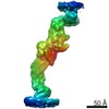
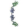
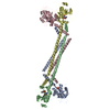
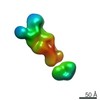
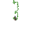
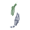
| Title | Munc13-1 C1-C2B-MUN-C2C Lateral conformation on lipid bilayer surface | |||||||||
 Components Components | Protein unc-13 homolog A | |||||||||
 Keywords Keywords | EXOCYTOSIS / Synaptic Transmission / Munc13 / Membrane Fusion | |||||||||
| Function / homology |  Function and homology information Function and homology informationdense core granule priming / neuronal dense core vesicle exocytosis / regulation of calcium-dependent activation of synaptic vesicle fusion / diacylglycerol binding / synaptic vesicle maturation / regulation of synaptic vesicle priming / : / positive regulation of vesicle fusion / positive regulation of dendrite extension / positive regulation of synaptic plasticity ...dense core granule priming / neuronal dense core vesicle exocytosis / regulation of calcium-dependent activation of synaptic vesicle fusion / diacylglycerol binding / synaptic vesicle maturation / regulation of synaptic vesicle priming / : / positive regulation of vesicle fusion / positive regulation of dendrite extension / positive regulation of synaptic plasticity / positive regulation of glutamate receptor signaling pathway / regulation of short-term neuronal synaptic plasticity / neurotransmitter secretion / regulation of amyloid precursor protein catabolic process / innervation / syntaxin binding / syntaxin-1 binding / presynaptic active zone / Golgi-associated vesicle / spectrin binding / positive regulation of neurotransmitter secretion / SNARE complex assembly / neuromuscular junction development / synaptic vesicle priming / amyloid-beta metabolic process / synaptic vesicle exocytosis / excitatory synapse / calyx of Held / SNARE binding / synaptic membrane / neuromuscular junction / synaptic transmission, glutamatergic / phospholipid binding / terminal bouton / long-term synaptic potentiation / synaptic vesicle membrane / presynapse / presynaptic membrane / cell differentiation / calmodulin binding / neuron projection / protein domain specific binding / axon / calcium ion binding / synapse / protein-containing complex binding / glutamatergic synapse / protein-containing complex / zinc ion binding / identical protein binding / plasma membrane Similarity search - Function | |||||||||
| Biological species |  | |||||||||
| Method | ELECTRON MICROSCOPY / subtomogram averaging / cryo EM / Resolution: 10 Å | |||||||||
 Authors Authors | Grushin, K. / Sindelar, C.V. | |||||||||
| Funding support |  United States, 2items United States, 2items
| |||||||||
 Citation Citation | Journal: Proc Natl Acad Sci U S A / Year: 2022 Title: Munc13 structural transitions and oligomers that may choreograph successive stages in vesicle priming for neurotransmitter release. Authors: Kirill Grushin / R Venkat Kalyana Sundaram / Charles V Sindelar / James E Rothman / Abstract: How can exactly six SNARE complexes be assembled under each synaptic vesicle? Here we report cryo-EM crystal structures of the core domain of Munc13, the key chaperone that initiates SNAREpin ...How can exactly six SNARE complexes be assembled under each synaptic vesicle? Here we report cryo-EM crystal structures of the core domain of Munc13, the key chaperone that initiates SNAREpin assembly. The functional core of Munc13, consisting of C1-C2B-MUN-C2C (Munc13C) spontaneously crystallizes between phosphatidylserine-rich bilayers in two distinct conformations, each in a radically different oligomeric state. In the open conformation (state 1), Munc13C forms upright trimers that link the two bilayers, separating them by ∼21 nm. In the closed conformation, six copies of Munc13C interact to form a lateral hexamer elevated ∼14 nm above the bilayer. Open and closed conformations differ only by a rigid body rotation around a flexible hinge, which when performed cooperatively assembles Munc13 into a lateral hexamer (state 2) in which the key SNARE assembly-activating site of Munc13 is autoinhibited by its neighbor. We propose that each Munc13 in the lateral hexamer ultimately assembles a single SNAREpin, explaining how only and exactly six SNARE complexes are templated. We suggest that state 1 and state 2 may represent two successive states in the synaptic vesicle supply chain leading to "primed" ready-release vesicles in which SNAREpins are clamped and ready to release (state 3). | |||||||||
| History |
|
- Structure visualization
| Movie |
Movie viewer |
|---|---|
| Structure viewer | Molecule: MolmilJmol/JSmol |
- Downloads & links
-Download
| PDBx/mmCIF format | 7t7v.cif.gz | 381.2 KB | Display | PDBx/mmCIF format |
|---|---|---|---|---|
| PDB format | pdb7t7v.ent.gz | 312 KB | Display | PDB format |
| PDBx/mmJSON format | 7t7v.json.gz | Tree view | PDBx/mmJSON format | |
| Others |  Other downloads Other downloads |
-Validation report
| Arichive directory | https://data.pdbj.org/pub/pdb/validation_reports/t7/7t7vftp://data.pdbj.org/pub/pdb/validation_reports/t7/7t7v | HTTPS FTP |
|---|
-Related structure data
| Related structure data |  25739MC  7t7cC  7t7rC  7t7xC  7t81C C: citing same article ( M: map data used to model this data |
|---|---|
| Similar structure data |


-Links
 PDBj
PDBj


- Assembly
| Deposited unit | 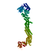
|
|---|---|
| 1 |
|
-Components
| #1: Protein | Mass: 130895.867 Da / Num. of mol.: 1 Source method: isolated from a genetically manipulated source Source: (gene. exp.)  Homo sapiens (human) / References: UniProt: Q4KUS2 Homo sapiens (human) / References: UniProt: Q4KUS2 |
|---|
-Experimental details
-Experiment
| Experiment | Method: ELECTRON MICROSCOPY |
|---|---|
| EM experiment | Aggregation state: 2D ARRAY / 3D reconstruction method: subtomogram averaging |
- Sample preparation
| Component | Name: 2D crystal of Munc13-1 C1-C2B-MUN-C2C domains between two lipid bilayers. Type: COMPLEX / Entity ID: all / Source: RECOMBINANT | ||||||||||||||||||||
|---|---|---|---|---|---|---|---|---|---|---|---|---|---|---|---|---|---|---|---|---|---|
| Molecular weight | Value: 0.13 MDa / Experimental value: NO | ||||||||||||||||||||
| Source (natural) | Organism: | ||||||||||||||||||||
| Source (recombinant) | Organism: Homo sapiens (human) / Cell: ExpiHEK-293 | ||||||||||||||||||||
| Buffer solution | pH: 7.4 | ||||||||||||||||||||
| Buffer component |
| ||||||||||||||||||||
| Specimen | Embedding applied: NO / Shadowing applied: NO / Staining applied: NO / Vitrification applied: YES | ||||||||||||||||||||
| Vitrification | Instrument: FEI VITROBOT MARK IV / Cryogen name: ETHANE / Humidity: 100 % / Chamber temperature: 281 K / Details: blot for 5 sec before plunging, blot force -1 |
- Electron microscopy imaging
| Experimental equipment |  Model: Titan Krios / Image courtesy: FEI Company |
|---|---|
| Microscopy | Model: FEI TITAN KRIOS |
| Electron gun | Electron source:  FIELD EMISSION GUN / Accelerating voltage: 300 kV / Illumination mode: FLOOD BEAM FIELD EMISSION GUN / Accelerating voltage: 300 kV / Illumination mode: FLOOD BEAM |
| Electron lens | Mode: BRIGHT FIELD / Nominal defocus max: 5000 nm / Nominal defocus min: 3500 nm |
| Image recording | Electron dose: 3.1 e/Å2 / Avg electron dose per subtomogram: 110 e/Å2 / Film or detector model: GATAN K3 (6k x 4k) |
| EM imaging optics | Energyfilter name: GIF Quantum LS / Energyfilter slit width: 20 eV |
- Processing
| EM software |
| ||||||||||||||||||||||||||||
|---|---|---|---|---|---|---|---|---|---|---|---|---|---|---|---|---|---|---|---|---|---|---|---|---|---|---|---|---|---|
| CTF correction | Details: CTF correction was performed during 3D reconstruction in RELION 3.1 Type: PHASE FLIPPING AND AMPLITUDE CORRECTION | ||||||||||||||||||||||||||||
| Symmetry | Point symmetry: C1 (asymmetric) | ||||||||||||||||||||||||||||
| 3D reconstruction | Resolution: 10 Å / Resolution method: OTHER / Num. of particles: 72894 Details: Combined map of best classes from two 3D classifications in RELION 3.1 of selected regions without angular searches using C6 symmetry expanded dataset. Num. of class averages: 8 / Symmetry type: POINT | ||||||||||||||||||||||||||||
| EM volume selection | Details: Particles were extracted and refined using Warp/M software Num. of tomograms: 62 / Num. of volumes extracted: 36837 | ||||||||||||||||||||||||||||
| Atomic model building | Protocol: FLEXIBLE FIT Details: Model for fitting was generated by AlphaFold using the construct's amino acid sequence. Flexible fitting into 3D map densities was performed using ISOLDE tool in ChimeraX. |
