 Movie
Movie Controller


+ Open data

- Basic information
| Entry | Database: PDB / ID: 7t20 | ||||||
|---|---|---|---|---|---|---|---|
| Title | E. coli DnaB bound to ssDNA and AMPPNP | ||||||
 Components Components |
| ||||||
 Keywords Keywords | REPLICATION/DNA / helicase / SF4 / AAA+ / REPLICATION-DNA complex | ||||||
| Function / homology |  Function and homology information Function and homology informationDnaB-DnaC complex / DnaB-DnaC-Rep-PriC complex / DnaB-DnaG complex / DnaB-DnaC-DnaT-PriA-PriC complex / DnaB-DnaC-DnaT-PriA-PriB complex / DNA helicase complex / primosome complex / DNA replication, synthesis of primer / DNA 5'-3' helicase / replisome ...DnaB-DnaC complex / DnaB-DnaC-Rep-PriC complex / DnaB-DnaG complex / DnaB-DnaC-DnaT-PriA-PriC complex / DnaB-DnaC-DnaT-PriA-PriB complex / DNA helicase complex / primosome complex / DNA replication, synthesis of primer / DNA 5'-3' helicase / replisome / response to ionizing radiation / replication fork processing / DNA replication initiation / DNA helicase activity / helicase activity / 5'-3' DNA helicase activity / DNA replication / ATP hydrolysis activity / DNA binding / ATP binding / identical protein binding / cytosol Similarity search - Function | ||||||
| Biological species |  | ||||||
| Method | ELECTRON MICROSCOPY / single particle reconstruction / cryo EM / Resolution: 4.7 Å | ||||||
 Authors Authors | Oakley, A.J. / Xu, Z.Q. | ||||||
| Funding support |  Australia, 1items Australia, 1items
| ||||||
 Citation Citation | Journal: To Be Published Title: Sequence of conformation changes of DnaB helicase during DNA unwinding and priming in Escherichia coli Authors: Oakley, A.J. / Xu, Z.Q. | ||||||
| History |
|
- Structure visualization
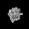
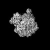
| Structure viewer | Molecule: MolmilJmol/JSmol |
|---|
- Downloads & links
-Download
| PDBx/mmCIF format | 7t20.cif.gz | 458.2 KB | Display | PDBx/mmCIF format |
|---|---|---|---|---|
| PDB format | pdb7t20.ent.gz | 380.3 KB | Display | PDB format |
| PDBx/mmJSON format | 7t20.json.gz | Tree view | PDBx/mmJSON format | |
| Others |  Other downloads Other downloads |
-Validation report
| Arichive directory | https://data.pdbj.org/pub/pdb/validation_reports/t2/7t20ftp://data.pdbj.org/pub/pdb/validation_reports/t2/7t20 | HTTPS FTP |
|---|
-Related structure data
| Related structure data | 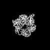 25607MC  7t21C  7t22C 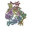 9ecoC M: map data used to model this data C: citing same article ( |
|---|---|
| Similar structure data |


-Links
 PDBj
PDBj


- Assembly
| Deposited unit | 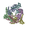
|
|---|---|
| 1 |
|
-Components
| #1: Protein | Mass: 52450.945 Da / Num. of mol.: 6 Source method: isolated from a genetically manipulated source Source: (gene. exp.) #2: DNA chain | | Mass: 6038.899 Da / Num. of mol.: 1 / Source method: obtained synthetically / Source: (synth.) #3: Chemical | ChemComp-ANP /   Mass: 506.196 Da / Num. of mol.: 4 / Source method: obtained synthetically / Formula: C10H17N6O12P3 / Feature type: SUBJECT OF INVESTIGATION / Comment: AMP-PNP, energy-carrying molecule analogue*YM Mass: 506.196 Da / Num. of mol.: 4 / Source method: obtained synthetically / Formula: C10H17N6O12P3 / Feature type: SUBJECT OF INVESTIGATION / Comment: AMP-PNP, energy-carrying molecule analogue*YM#4: Chemical | ChemComp-MG /   Mass: 24.305 Da / Num. of mol.: 4 / Source method: obtained synthetically / Formula: Mg / Feature type: SUBJECT OF INVESTIGATION Mass: 24.305 Da / Num. of mol.: 4 / Source method: obtained synthetically / Formula: Mg / Feature type: SUBJECT OF INVESTIGATIONHas ligand of interest | Y | |
|---|
-Experimental details
-Experiment
| Experiment | Method: ELECTRON MICROSCOPY |
|---|---|
| EM experiment | Aggregation state: PARTICLE / 3D reconstruction method: single particle reconstruction |
- Sample preparation
| Component | Name: E. coli DnaB bound to ssDNA / Type: COMPLEX / Entity ID: #1-#2 / Source: MULTIPLE SOURCES | ||||||||||||
|---|---|---|---|---|---|---|---|---|---|---|---|---|---|
| Molecular weight | Units: KILODALTONS/NANOMETER / Experimental value: YES | ||||||||||||
| Source (natural) |
| ||||||||||||
| Source (recombinant) | Organism: | ||||||||||||
| Buffer solution | pH: 7.6 Details: 20 mM Tris-HCl, pH 7.6, 100 mM NaCl, 5 mM MgCl2, 3 mM DTT, 0.25 mM EDTA and 100 micromolar ADP. | ||||||||||||
| Specimen | Conc.: 0.5 mg/ml / Embedding applied: NO / Shadowing applied: NO / Staining applied: NO / Vitrification applied: YES | ||||||||||||
| Specimen support | Details: Grids covered with a 2 nm continuous carbon film. Glow-discharged grids were floated on 40 microL drops of a 0.1% (w/v) aqueous solution of poly-lysine hydrobromide (MW 30-70 kDa; ...Details: Grids covered with a 2 nm continuous carbon film. Glow-discharged grids were floated on 40 microL drops of a 0.1% (w/v) aqueous solution of poly-lysine hydrobromide (MW 30-70 kDa; Polysciences, Inc.) for two min, briefly rinsed with MilliQ water 4 times and then allow to air dry. Grid material: COPPER / Grid mesh size: 300 divisions/in. / Grid type: Quantifoil R1.2/1.3 | ||||||||||||
| Vitrification | Instrument: FEI VITROBOT MARK IV / Cryogen name: ETHANE / Chamber temperature: 279 K Details: 3 microL of sample was applied to glow-discharged grids. Grids were blotted at 6 degrees C for 3.5 s with no extra blot force. |
- Electron microscopy imaging
| Experimental equipment |  Model: Talos Arctica / Image courtesy: FEI Company |
|---|---|
| Microscopy | Model: FEI TALOS ARCTICA |
| Electron gun | Electron source:  FIELD EMISSION GUN / Accelerating voltage: 200 kV / Illumination mode: FLOOD BEAM FIELD EMISSION GUN / Accelerating voltage: 200 kV / Illumination mode: FLOOD BEAM |
| Electron lens | Mode: BRIGHT FIELD |
| Specimen holder | Cryogen: NITROGEN / Specimen holder model: FEI TITAN KRIOS AUTOGRID HOLDER |
| Image recording | Electron dose: 50 e/Å2 / Detector mode: COUNTING / Film or detector model: FEI FALCON III (4k x 4k) |
- Processing
| Software | Name: PHENIX / Version: dev_4370: / Classification: refinement | ||||||||||||||||||||||||||||||||||||||||
|---|---|---|---|---|---|---|---|---|---|---|---|---|---|---|---|---|---|---|---|---|---|---|---|---|---|---|---|---|---|---|---|---|---|---|---|---|---|---|---|---|---|
| EM software |
| ||||||||||||||||||||||||||||||||||||||||
| CTF correction | Type: PHASE FLIPPING AND AMPLITUDE CORRECTION | ||||||||||||||||||||||||||||||||||||||||
| Particle selection | Num. of particles selected: 1098240 | ||||||||||||||||||||||||||||||||||||||||
| Symmetry | Point symmetry: C1 (asymmetric) | ||||||||||||||||||||||||||||||||||||||||
| 3D reconstruction | Resolution: 4.7 Å / Resolution method: FSC 0.143 CUT-OFF / Num. of particles: 373620 / Algorithm: FOURIER SPACE / Num. of class averages: 1 / Symmetry type: POINT | ||||||||||||||||||||||||||||||||||||||||
| Atomic model building | Protocol: OTHER / Space: REAL / Target criteria: Correlation Coefficient | ||||||||||||||||||||||||||||||||||||||||
| Refine LS restraints |
|
