Movie
Movie Controller

[English] 日本語
 Yorodumi
- PDB-7syh: Structure of the HCV IRES binding to the 40S ribosomal subunit, c... -
+ Open data

- Basic information
| Entry | Database: PDB / ID: 7syh | |||||||||||||||
|---|---|---|---|---|---|---|---|---|---|---|---|---|---|---|---|---|
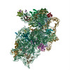
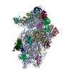
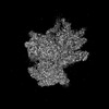
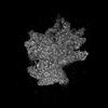
| Title | Structure of the HCV IRES binding to the 40S ribosomal subunit, closed conformation. Structure 2(delta dII) | |||||||||||||||
 Components Components |
| |||||||||||||||
 Keywords Keywords | RIBOSOME / HCV / IRES / 40S | |||||||||||||||
| Function / homology |  Function and homology information Function and homology informationcytoplasmic side of rough endoplasmic reticulum membrane / laminin receptor activity / 90S preribosome / ubiquitin ligase inhibitor activity / positive regulation of signal transduction by p53 class mediator / phagocytic cup / translation regulator activity / rough endoplasmic reticulum / ribosomal small subunit export from nucleus / laminin binding ...cytoplasmic side of rough endoplasmic reticulum membrane / laminin receptor activity / 90S preribosome / ubiquitin ligase inhibitor activity / positive regulation of signal transduction by p53 class mediator / phagocytic cup / translation regulator activity / rough endoplasmic reticulum / ribosomal small subunit export from nucleus / laminin binding / gastrulation / MDM2/MDM4 family protein binding / cytosolic ribosome / class I DNA-(apurinic or apyrimidinic site) endonuclease activity / DNA-(apurinic or apyrimidinic site) lyase / maturation of SSU-rRNA from tricistronic rRNA transcript (SSU-rRNA, 5.8S rRNA, LSU-rRNA) / positive regulation of apoptotic signaling pathway / maturation of SSU-rRNA / small-subunit processome / spindle / rRNA processing / positive regulation of canonical Wnt signaling pathway / rhythmic process / regulation of translation / ribosomal small subunit assembly / virus receptor activity / ribosome binding / ribosomal small subunit biogenesis / small ribosomal subunit / small ribosomal subunit rRNA binding / cytosolic small ribosomal subunit / perikaryon / cell differentiation / cytoplasmic translation / mitochondrial inner membrane / postsynaptic density / rRNA binding / structural constituent of ribosome / ribosome / translation / ribonucleoprotein complex / cell division / DNA repair / mRNA binding / apoptotic process / centrosome / synapse / dendrite / nucleolus / perinuclear region of cytoplasm / Golgi apparatus / DNA binding / RNA binding / zinc ion binding / membrane / nucleus / plasma membrane / cytoplasm Similarity search - Function | |||||||||||||||
| Biological species |  Hepatitis C virus Hepatitis C virus | |||||||||||||||
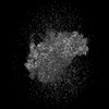
| Method | ELECTRON MICROSCOPY / single particle reconstruction / cryo EM / Resolution: 4.6 Å | |||||||||||||||
 Authors Authors | Brown, Z.P. / Abaeva, I.S. / De, S. / Hellen, C.U.T. / Pestova, T.V. / Frank, J. | |||||||||||||||
| Funding support |  United States, 4items United States, 4items
| |||||||||||||||
 Citation Citation | Journal: To Be Published Title: Molecular architecture of 40S initiation complexes on the Hepatitis C virus IRES: from ribosomal attachment to eIF5B-mediated reorientation of initiator tRNA Authors: Brown, Z.P. / Abaeva, I.S. / De, S. / Hellen, C.U.T. / Pestova, T.V. / Frank, J. | |||||||||||||||
| History |
|
- Structure visualization
| Structure viewer | Molecule: MolmilJmol/JSmol |
|---|
- Downloads & links
-Download
| PDBx/mmCIF format | 7syh.cif.gz | 1.7 MB | Display | PDBx/mmCIF format |
|---|---|---|---|---|
| PDB format | pdb7syh.ent.gz | 1.3 MB | Display | PDB format |
| PDBx/mmJSON format | 7syh.json.gz | Tree view | PDBx/mmJSON format | |
| Others |  Other downloads Other downloads |
-Validation report
| Arichive directory | https://data.pdbj.org/pub/pdb/validation_reports/sy/7syhftp://data.pdbj.org/pub/pdb/validation_reports/sy/7syh | HTTPS FTP |
|---|
-Related structure data
| Related structure data | 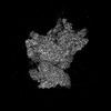 25528MC  7sygC  7synC M: map data used to model this data C: citing same article ( |
|---|---|
| Similar structure data |

-Links
 PDBj
PDBj


- Assembly
| Deposited unit | 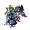
|
|---|---|
| 1 |
|
-Components
-RNA chain , 2 types, 2 molecules 2z
| #1: RNA chain | Mass: 603100.938 Da / Num. of mol.: 1 / Source method: isolated from a natural source / Source: (natural) |
|---|---|
| #36: RNA chain | Mass: 128746.109 Da / Num. of mol.: 1 Source method: isolated from a genetically manipulated source Source: (gene. exp.) Hepatitis C virus (isolate 1) / Production host:  |
-40S ribosomal protein ... , 9 types, 9 molecules BCFIWZabg
| #2: Protein | Mass: 32958.016 Da / Num. of mol.: 1 / Source method: isolated from a natural source / Source: (natural) |
|---|---|
| #3: Protein | Mass: 30002.061 Da / Num. of mol.: 1 / Source method: isolated from a natural source / Source: (natural) |
| #6: Protein | Mass: 29654.869 Da / Num. of mol.: 1 / Source method: isolated from a natural source / Source: (natural) |
| #9: Protein | Mass: 47356.172 Da / Num. of mol.: 1 / Source method: isolated from a natural source / Source: (natural) |
| #23: Protein | Mass: 9124.389 Da / Num. of mol.: 1 / Source method: isolated from a natural source / Source: (natural) |
| #26: Protein | Mass: 15237.104 Da / Num. of mol.: 1 / Source method: isolated from a natural source / Source: (natural) |
| #27: Protein | Mass: 13645.028 Da / Num. of mol.: 1 / Source method: isolated from a natural source / Source: (natural) |
| #28: Protein | Mass: 11645.794 Da / Num. of mol.: 1 / Source method: isolated from a natural source / Source: (natural) |
| #33: Protein | Mass: 21431.742 Da / Num. of mol.: 1 / Source method: isolated from a natural source / Source: (natural) |
+Protein , 24 types, 24 molecules DEGHJKLMNOPQRSTUVXYcdefh
-Protein/peptide / Non-polymers , 2 types, 3 molecules n
| #35: Protein/peptide | Mass: 3473.451 Da / Num. of mol.: 1 / Source method: isolated from a natural source / Source: (natural) |
|---|---|
| #37: Chemical |  Mass: 65.409 Da / Num. of mol.: 2 / Source method: obtained synthetically / Formula: Zn Mass: 65.409 Da / Num. of mol.: 2 / Source method: obtained synthetically / Formula: Zn |
-Details
| Has ligand of interest | N |
|---|---|
| Has protein modification | Y |
-Experimental details
-Experiment
| Experiment | Method: ELECTRON MICROSCOPY |
|---|---|
| EM experiment | Aggregation state: PARTICLE / 3D reconstruction method: single particle reconstruction |
- Sample preparation
| Component | Name: 40S ribosomal small subunit with HCV IRES / Type: RIBOSOME / Entity ID: #1-#27, #29-#36 / Source: MULTIPLE SOURCES | ||||||||||||
|---|---|---|---|---|---|---|---|---|---|---|---|---|---|
| Molecular weight | Value: 2 MDa / Experimental value: NO | ||||||||||||
| Source (natural) |
| ||||||||||||
| Source (recombinant) | Organism: | ||||||||||||
| Buffer solution | pH: 7.5 | ||||||||||||
| Specimen | Conc.: 7.5E-5 mg/ml / Embedding applied: NO / Shadowing applied: NO / Staining applied: NO / Vitrification applied: YES | ||||||||||||
| Specimen support | Details: H2/O2 mixture for 25 seconds at 25W power / Grid material: GOLD / Grid mesh size: 300 divisions/in. / Grid type: Quantifoil R0.6/1 | ||||||||||||
| Vitrification | Instrument: FEI VITROBOT MARK IV / Cryogen name: ETHANE-PROPANE / Humidity: 100 % / Chamber temperature: 277.15 K / Details: 4 second blot time, force 3 |
- Electron microscopy imaging
| Experimental equipment |  Model: Tecnai F30 / Image courtesy: FEI Company |
|---|---|
| Microscopy | Model: FEI TECNAI F30 |
| Electron gun | Electron source:  FIELD EMISSION GUN / Accelerating voltage: 300 kV / Illumination mode: FLOOD BEAM FIELD EMISSION GUN / Accelerating voltage: 300 kV / Illumination mode: FLOOD BEAM |
| Electron lens | Mode: BRIGHT FIELD / Nominal magnification: 52000 X / Nominal defocus max: 2000 nm / Nominal defocus min: 1000 nm / Cs: 2.26 mm / Alignment procedure: COMA FREE |
| Specimen holder | Cryogen: NITROGEN |
| Image recording | Average exposure time: 4 sec. / Electron dose: 70.9 e/Å2 / Film or detector model: GATAN K3 (6k x 4k) |
| Image scans | Sampling size: 5 µm |
- Processing
| Software | Name: PHENIX / Version: 1.19.1_4122: / Classification: refinement | ||||||||||||||||||||||||||||||||
|---|---|---|---|---|---|---|---|---|---|---|---|---|---|---|---|---|---|---|---|---|---|---|---|---|---|---|---|---|---|---|---|---|---|
| EM software |
| ||||||||||||||||||||||||||||||||
| CTF correction | Type: PHASE FLIPPING AND AMPLITUDE CORRECTION | ||||||||||||||||||||||||||||||||
| Symmetry | Point symmetry: C1 (asymmetric) | ||||||||||||||||||||||||||||||||
| 3D reconstruction | Resolution: 4.6 Å / Resolution method: FSC 0.143 CUT-OFF / Num. of particles: 28684 / Symmetry type: POINT | ||||||||||||||||||||||||||||||||
| Atomic model building | 3D fitting-ID: 1 / Source name: PDB / Type: experimental model
| ||||||||||||||||||||||||||||||||
| Refine LS restraints |
|