 Movie
Movie Controller


+ Open data

- Basic information
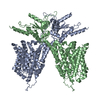
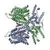
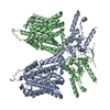
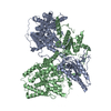
| Entry | Database: PDB / ID: 7sun | ||||||
|---|---|---|---|---|---|---|---|
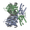
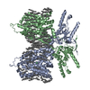
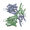
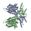
| Title | Atomic model of prestin from gerbil (Meriones unguiculatus) | ||||||
 Components Components | Prestin, Enhanced Yellow Fluorescent Protein chimera | ||||||
 Keywords Keywords | MEMBRANE PROTEIN / MOTOR PROTEIN / Prestin (Slc26a5) / cochlear amplification / NonLinear Capacitance | ||||||
| Function / homology |  Function and homology information Function and homology informationsecondary active sulfate transmembrane transporter activity / chloride:bicarbonate antiporter activity / bicarbonate transport / chloride transport / lateral plasma membrane / sensory perception of sound / regulation of cell shape / basolateral plasma membrane / protein homodimerization activity Similarity search - Function | ||||||
| Biological species |  Meriones unguiculatus (Mongolian gerbil) Meriones unguiculatus (Mongolian gerbil)synthetic construct (others) | ||||||
| Method | ELECTRON MICROSCOPY / single particle reconstruction / cryo EM / Resolution: 3.6 Å | ||||||
 Authors Authors | Butan, C. / Santos-Sacchi, J. | ||||||
| Funding support |  United States, 1items United States, 1items
| ||||||
 Citation Citation | Journal: Nat Commun / Year: 2022 Title: Single particle cryo-EM structure of the outer hair cell motor protein prestin. Authors: Carmen Butan / Qiang Song / Jun-Ping Bai / Winston J T Tan / Dhasakumar Navaratnam / Joseph Santos-Sacchi / Abstract: The mammalian outer hair cell (OHC) protein prestin (Slc26a5) differs from other Slc26 family members due to its unique piezoelectric-like property that drives OHC electromotility, the putative ...The mammalian outer hair cell (OHC) protein prestin (Slc26a5) differs from other Slc26 family members due to its unique piezoelectric-like property that drives OHC electromotility, the putative mechanism for cochlear amplification. Here, we use cryo-electron microscopy to determine prestin's structure at 3.6 Å resolution. Prestin is structurally similar to the anion transporter Slc26a9. It is captured in an inward-open state which may reflect prestin's contracted state. Two well-separated transmembrane (TM) domains and two cytoplasmic sulfate transporter and anti-sigma factor antagonist (STAS) domains form a swapped dimer. The transmembrane domains consist of 14 transmembrane segments organized in two 7+7 inverted repeats, an architecture first observed in the bacterial symporter UraA. Mutation of prestin's chloride binding site removes salicylate competition with anions while retaining the prestin characteristic displacement currents (Nonlinear Capacitance), undermining the extrinsic voltage sensor hypothesis for prestin function. | ||||||
| History |
|
- Structure visualization
| Movie |
Movie viewer |
|---|---|
| Structure viewer | Molecule: MolmilJmol/JSmol |
- Downloads & links
-Download
| PDBx/mmCIF format | 7sun.cif.gz | 247.1 KB | Display | PDBx/mmCIF format |
|---|---|---|---|---|
| PDB format | pdb7sun.ent.gz | 192.2 KB | Display | PDB format |
| PDBx/mmJSON format | 7sun.json.gz | Tree view | PDBx/mmJSON format | |
| Others |  Other downloads Other downloads |
-Validation report
| Arichive directory | https://data.pdbj.org/pub/pdb/validation_reports/su/7sunftp://data.pdbj.org/pub/pdb/validation_reports/su/7sun | HTTPS FTP |
|---|
-Related structure data
| Related structure data | 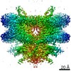 25442MC M: map data used to model this data C: citing same article ( |
|---|---|
| Similar structure data |


-Links
 PDBj
PDBj
- Assembly
| Deposited unit | 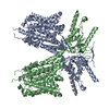
|
|---|---|
| 1 |
|
-Components
| #1: Protein | Mass: 111865.805 Da / Num. of mol.: 2 / Fragment: prestin + linker + eYFP Source method: isolated from a genetically manipulated source Source: (gene. exp.) Meriones unguiculatus (Mongolian gerbil), (gene. exp.) synthetic construct (others)Gene: SLC26A5, PRES, eYFP / Cell line (production host): HEK293 / Production host:  Homo sapiens (human) / References: UniProt: Q9JKQ2 Homo sapiens (human) / References: UniProt: Q9JKQ2 |
|---|
-Experimental details
-Experiment
| Experiment | Method: ELECTRON MICROSCOPY |
|---|---|
| EM experiment | Aggregation state: PARTICLE / 3D reconstruction method: single particle reconstruction |
- Sample preparation
| Component | Name: Prestin dimer surrounded by a belt of detergent / Type: COMPLEX / Entity ID: all / Source: RECOMBINANT |
|---|---|
| Source (natural) | Organism: Meriones unguiculatus (Mongolian gerbil) |
| Source (recombinant) | Organism: Homo sapiens (human) / Cell: HEK293 |
| Buffer solution | pH: 7.4 / Details: 10 mM HEPES, 200 mM NaCl, 0.02% GDN, pH 7.4 |
| Specimen | Embedding applied: NO / Shadowing applied: NO / Staining applied: NO / Vitrification applied: YES |
| Vitrification | Cryogen name: ETHANE |
- Electron microscopy imaging
| Experimental equipment |  Model: Titan Krios / Image courtesy: FEI Company |
|---|---|
| Microscopy | Model: TFS KRIOS |
| Electron gun | Electron source:  FIELD EMISSION GUN / Accelerating voltage: 300 kV / Illumination mode: FLOOD BEAM FIELD EMISSION GUN / Accelerating voltage: 300 kV / Illumination mode: FLOOD BEAM |
| Electron lens | Mode: BRIGHT FIELD / Nominal magnification: 81000 X / Nominal defocus max: 2150 nm / Nominal defocus min: 1150 nm |
| Image recording | Electron dose: 54 e/Å2 / Film or detector model: GATAN K3 (6k x 4k) |
- Processing
| Software | Name: PHENIX / Version: 1.19.2_4158: / Classification: refinement | ||||||||||||||||||||||||||||
|---|---|---|---|---|---|---|---|---|---|---|---|---|---|---|---|---|---|---|---|---|---|---|---|---|---|---|---|---|---|
| EM software |
| ||||||||||||||||||||||||||||
| CTF correction | Type: PHASE FLIPPING AND AMPLITUDE CORRECTION | ||||||||||||||||||||||||||||
| Symmetry | Point symmetry: C2 (2 fold cyclic) | ||||||||||||||||||||||||||||
| 3D reconstruction | Resolution: 3.6 Å / Resolution method: FSC 0.143 CUT-OFF / Num. of particles: 111863 / Symmetry type: POINT | ||||||||||||||||||||||||||||
| Atomic model building | PDB-ID: 6RTF Accession code: 6RTF / Source name: PDB / Type: experimental model | ||||||||||||||||||||||||||||
| Refine LS restraints |
|

