 Movie
Movie Controller


+ Open data

- Basic information
| Entry | Database: PDB / ID: 7sc5 | ||||||
|---|---|---|---|---|---|---|---|
| Title | Cytoplasmic tail deleted HIV Env trimer in nanodisc | ||||||
 Components Components |
| ||||||
 Keywords Keywords | VIRAL PROTEIN / HIV / Spike / trimer / nanodisc | ||||||
| Function / homology |  Function and homology information Function and homology informationpositive regulation of plasma membrane raft polarization / positive regulation of receptor clustering / positive regulation of establishment of T cell polarity / host cell endosome membrane / clathrin-dependent endocytosis of virus by host cell / viral protein processing / fusion of virus membrane with host plasma membrane / virus-mediated perturbation of host defense response / fusion of virus membrane with host endosome membrane / viral envelope ...positive regulation of plasma membrane raft polarization / positive regulation of receptor clustering / positive regulation of establishment of T cell polarity / host cell endosome membrane / clathrin-dependent endocytosis of virus by host cell / viral protein processing / fusion of virus membrane with host plasma membrane / virus-mediated perturbation of host defense response / fusion of virus membrane with host endosome membrane / viral envelope / virion attachment to host cell / apoptotic process / host cell plasma membrane / structural molecule activity / virion membrane / plasma membrane Similarity search - Function | ||||||
| Biological species | HIV whole-genome vector AA1305#18 (others) | ||||||
| Method | ELECTRON MICROSCOPY / single particle reconstruction / cryo EM / Resolution: 3.88 Å | ||||||
 Authors Authors | Yang, S. / Walz, T. | ||||||
| Funding support |  United States, 1items United States, 1items
| ||||||
 Citation Citation | Journal: Nat Commun / Year: 2022 Title: Dynamic HIV-1 spike motion creates vulnerability for its membrane-bound tripod to antibody attack. Authors: Shuang Yang / Giorgos Hiotis / Yi Wang / Junjian Chen / Jia-Huai Wang / Mikyung Kim / Ellis L Reinherz / Thomas Walz / Abstract: Vaccines targeting HIV-1's gp160 spike protein are stymied by high viral mutation rates and structural chicanery. gp160's membrane-proximal external region (MPER) is the target of naturally arising ...Vaccines targeting HIV-1's gp160 spike protein are stymied by high viral mutation rates and structural chicanery. gp160's membrane-proximal external region (MPER) is the target of naturally arising broadly neutralizing antibodies (bnAbs), yet MPER-based vaccines fail to generate bnAbs. Here, nanodisc-embedded spike protein was investigated by cryo-electron microscopy and molecular-dynamics simulations, revealing spontaneous ectodomain tilting that creates vulnerability for HIV-1. While each MPER protomer radiates centrally towards the three-fold axis contributing to a membrane-associated tripod structure that is occluded in the upright spike, tilting provides access to the opposing MPER. Structures of spike proteins with bound 4E10 bnAb Fabs reveal that the antibody binds exposed MPER, thereby altering MPER dynamics, modifying average ectodomain tilt, and imposing strain on the viral membrane and the spike's transmembrane segments, resulting in the abrogation of membrane fusion and informing future vaccine development. | ||||||
| History |
|
- Structure visualization
| Structure viewer | Molecule: MolmilJmol/JSmol |
|---|
- Downloads & links
-Download
| PDBx/mmCIF format | 7sc5.cif.gz | 327.4 KB | Display | PDBx/mmCIF format |
|---|---|---|---|---|
| PDB format | pdb7sc5.ent.gz | 274.6 KB | Display | PDB format |
| PDBx/mmJSON format | 7sc5.json.gz | Tree view | PDBx/mmJSON format | |
| Others |  Other downloads Other downloads |
-Validation report
| Summary document | 7sc5_validation.pdf.gz | 2.3 MB | Display | wwPDB validaton report |
|---|---|---|---|---|
| Full document | 7sc5_full_validation.pdf.gz | 2.4 MB | Display | |
| Data in XML | 7sc5_validation.xml.gz | 59.2 KB | Display | |
| Data in CIF | 7sc5_validation.cif.gz | 84.7 KB | Display | |
| Arichive directory | https://data.pdbj.org/pub/pdb/validation_reports/sc/7sc5ftp://data.pdbj.org/pub/pdb/validation_reports/sc/7sc5 | HTTPS FTP |
-Related structure data
| Related structure data | 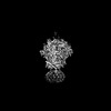 25022MC  7sd3C M: map data used to model this data C: citing same article ( |
|---|---|
| Similar structure data |


-Links
 PDBj
PDBj


- Assembly
| Deposited unit | 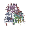
|
|---|---|
| 1 |
|
-Components
| #1: Protein | Mass: 53092.070 Da / Num. of mol.: 3 / Fragment: UNP residues 30-503 Source method: isolated from a genetically manipulated source Source: (gene. exp.) HIV whole-genome vector AA1305#18 (others) Gene: env / Cell line (production host): HEK293F / Production host:  Homo sapiens (human) / References: UniProt: A0A6H1VID3 Homo sapiens (human) / References: UniProt: A0A6H1VID3#2: Protein | Mass: 24114.770 Da / Num. of mol.: 3 / Fragment: UNP residues 509-720 Source method: isolated from a genetically manipulated source Source: (gene. exp.) HIV whole-genome vector AA1305#18 (others) Gene: env / Cell line (production host): HEK293F / Production host: Homo sapiens (human) / References: UniProt: A0A173DX29#3: Polysaccharide | beta-D-mannopyranose-(1-4)-2-acetamido-2-deoxy-beta-D-glucopyranose-(1-4)-2-acetamido-2-deoxy-beta- ...beta-D-mannopyranose-(1-4)-2-acetamido-2-deoxy-beta-D-glucopyranose-(1-4)-2-acetamido-2-deoxy-beta-D-glucopyranose Source method: isolated from a genetically manipulated source #4: Polysaccharide | Source method: isolated from a genetically manipulated source #5: Sugar | ChemComp-NAG /   Type: D-saccharide, beta linking / Mass: 221.208 Da / Num. of mol.: 36 / Source method: obtained synthetically / Formula: C8H15NO6 / Feature type: SUBJECT OF INVESTIGATION Type: D-saccharide, beta linking / Mass: 221.208 Da / Num. of mol.: 36 / Source method: obtained synthetically / Formula: C8H15NO6 / Feature type: SUBJECT OF INVESTIGATIONHas ligand of interest | Y | |
|---|
-Experimental details
-Experiment
| Experiment | Method: ELECTRON MICROSCOPY |
|---|---|
| EM experiment | Aggregation state: PARTICLE / 3D reconstruction method: single particle reconstruction |
- Sample preparation
| Component | Name: HIV-1 envelope glycoprotein Env trimer / Type: ORGANELLE OR CELLULAR COMPONENT / Entity ID: #1-#2 / Source: RECOMBINANT |
|---|---|
| Source (natural) | Organism: HIV whole-genome vector AA1305#18 (others) / Strain: BG505 |
| Source (recombinant) | Organism: Homo sapiens (human) / Cell: HEK293F |
| Buffer solution | pH: 7.5 |
| Specimen | Conc.: 0.2 mg/ml / Embedding applied: NO / Shadowing applied: NO / Staining applied: NO / Vitrification applied: YES |
| Specimen support | Grid material: GRAPHENE OXIDE / Grid type: Quantifoil R1.2/1.3 |
| Vitrification | Instrument: FEI VITROBOT MARK IV / Cryogen name: ETHANE / Humidity: 100 % / Chamber temperature: 277 K |
- Electron microscopy imaging
| Experimental equipment |  Model: Titan Krios / Image courtesy: FEI Company |
|---|---|
| Microscopy | Model: FEI TITAN KRIOS |
| Electron gun | Electron source:  FIELD EMISSION GUN / Accelerating voltage: 300 kV / Illumination mode: FLOOD BEAM FIELD EMISSION GUN / Accelerating voltage: 300 kV / Illumination mode: FLOOD BEAM |
| Electron lens | Mode: BRIGHT FIELD / Nominal defocus max: 2500 nm / Nominal defocus min: 1500 nm |
| Image recording | Electron dose: 80 e/Å2 / Film or detector model: GATAN K2 SUMMIT (4k x 4k) |
- Processing
| CTF correction | Type: NONE |
|---|---|
| 3D reconstruction | Resolution: 3.88 Å / Resolution method: FSC 0.143 CUT-OFF / Num. of particles: 47616 / Symmetry type: POINT |
