 Movie
Movie Controller


+ Open data

- Basic information
| Entry | Database: PDB / ID: 7sba | ||||||
|---|---|---|---|---|---|---|---|
| Title | Structure of type I-D Cascade bound to a dsDNA target | ||||||
 Components Components |
| ||||||
 Keywords Keywords | DNA BINDING PROTEIN/DNA/RNA / CRISPR / Complex / Ribonucleoprotein complex / type I-D / type ID / type I / cyanobacteria / synechocystis / RNA binding protein / DNA binding protein / DNA BINDING PROTEIN-DNA-RNA complex | ||||||
| Function / homology |  Function and homology information Function and homology informationCRISPR-associated protein Csc1 / CRISPR associated protein Csc3 / Type I-D CRISPR-associated protein Cas5/Csc1 / CRISPR-associated protein Csc2 / Csc2 Crispr Similarity search - Domain/homology | ||||||
| Biological species |  synthetic construct (others) | ||||||
| Method | ELECTRON MICROSCOPY / single particle reconstruction / cryo EM / Resolution: 2.9 Å | ||||||
 Authors Authors | Schwartz, E.A. / Taylor, D.W. | ||||||
| Funding support |  United States, 1items United States, 1items
| ||||||
 Citation Citation | Journal: Nat Commun / Year: 2022 Title: Structural rearrangements allow nucleic acid discrimination by type I-D Cascade. Authors: Evan A Schwartz / Tess M McBride / Jack P K Bravo / Daniel Wrapp / Peter C Fineran / Robert D Fagerlund / David W Taylor /  Abstract: CRISPR-Cas systems are adaptive immune systems that protect prokaryotes from foreign nucleic acids, such as bacteriophages. Two of the most prevalent CRISPR-Cas systems include type I and type III. ...CRISPR-Cas systems are adaptive immune systems that protect prokaryotes from foreign nucleic acids, such as bacteriophages. Two of the most prevalent CRISPR-Cas systems include type I and type III. Interestingly, the type I-D interference proteins contain characteristic features of both type I and type III systems. Here, we present the structures of type I-D Cascade bound to both a double-stranded (ds)DNA and a single-stranded (ss)RNA target at 2.9 and 3.1 Å, respectively. We show that type I-D Cascade is capable of specifically binding ssRNA and reveal how PAM recognition of dsDNA targets initiates long-range structural rearrangements that likely primes Cas10d for Cas3' binding and subsequent non-target strand DNA cleavage. These structures allow us to model how binding of the anti-CRISPR protein AcrID1 likely blocks target dsDNA binding via competitive inhibition of the DNA substrate engagement with the Cas10d active site. This work elucidates the unique mechanisms used by type I-D Cascade for discrimination of single-stranded and double stranded targets. Thus, our data supports a model for the hybrid nature of this complex with features of type III and type I systems. | ||||||
| History |
|
- Structure visualization
| Structure viewer | Molecule: MolmilJmol/JSmol |
|---|
- Downloads & links
-Download
| PDBx/mmCIF format | 7sba.cif.gz | 667.9 KB | Display | PDBx/mmCIF format |
|---|---|---|---|---|
| PDB format | pdb7sba.ent.gz | 547.7 KB | Display | PDB format |
| PDBx/mmJSON format | 7sba.json.gz | Tree view | PDBx/mmJSON format | |
| Others |  Other downloads Other downloads |
-Validation report
| Arichive directory | https://data.pdbj.org/pub/pdb/validation_reports/sb/7sbaftp://data.pdbj.org/pub/pdb/validation_reports/sb/7sba | HTTPS FTP |
|---|
-Related structure data
| Related structure data | 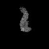 24974MC  7sbbC M: map data used to model this data C: citing same article ( |
|---|---|
| Similar structure data |

-Links
 PDBj
PDBj


- Assembly
| Deposited unit | 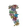
|
|---|---|
| 1 |
|
-Components
-Protein , 4 types, 11 molecules ABCDEFGHIJK
| #1: Protein | Mass: 36527.109 Da / Num. of mol.: 7 Source method: isolated from a genetically manipulated source Source: (gene. exp.) #2: Protein | | Mass: 28942.748 Da / Num. of mol.: 1 Source method: isolated from a genetically manipulated source Source: (gene. exp.) #3: Protein | | Mass: 112067.508 Da / Num. of mol.: 1 Source method: isolated from a genetically manipulated source Source: (gene. exp.) #4: Protein | Mass: 17024.494 Da / Num. of mol.: 2 Source method: isolated from a genetically manipulated source Source: (gene. exp.) |
|---|
-DNA chain , 2 types, 2 molecules XY
| #5: DNA chain | Mass: 4884.222 Da / Num. of mol.: 1 / Source method: obtained synthetically / Source: (synth.) synthetic construct (others) |
|---|---|
| #6: DNA chain | Mass: 3947.581 Da / Num. of mol.: 1 / Source method: obtained synthetically / Source: (synth.) synthetic construct (others) |
-RNA chain , 1 types, 1 molecules Z
| #7: RNA chain | Mass: 13699.081 Da / Num. of mol.: 1 Source method: isolated from a genetically manipulated source Source: (gene. exp.) |
|---|
-Experimental details
-Experiment
| Experiment | Method: ELECTRON MICROSCOPY |
|---|---|
| EM experiment | Aggregation state: PARTICLE / 3D reconstruction method: single particle reconstruction |
- Sample preparation
| Component | Name: Type I-D Cascade bound to a dsDNA target / Type: COMPLEX / Entity ID: all / Source: MULTIPLE SOURCES | |||||||||||||||||||||||||
|---|---|---|---|---|---|---|---|---|---|---|---|---|---|---|---|---|---|---|---|---|---|---|---|---|---|---|
| Molecular weight | Value: 0.48 MDa / Experimental value: NO | |||||||||||||||||||||||||
| Buffer solution | pH: 7.5 | |||||||||||||||||||||||||
| Buffer component |
| |||||||||||||||||||||||||
| Specimen | Conc.: 0.125 mg/ml / Embedding applied: NO / Shadowing applied: NO / Staining applied: NO / Vitrification applied: YES / Details: Monodisperse sample of elongated particles. | |||||||||||||||||||||||||
| Specimen support | Grid material: COPPER / Grid mesh size: 400 divisions/in. / Grid type: C-flat | |||||||||||||||||||||||||
| Vitrification | Instrument: FEI VITROBOT MARK IV / Cryogen name: ETHANE / Humidity: 100 % / Chamber temperature: 277 K Details: Blot force 0, blot time 4.5s, no wait time between sample application and blotting. |
- Electron microscopy imaging
| Experimental equipment |  Model: Titan Krios / Image courtesy: FEI Company |
|---|---|
| Microscopy | Model: FEI TITAN KRIOS |
| Electron gun | Electron source:  FIELD EMISSION GUN / Accelerating voltage: 300 kV / Illumination mode: FLOOD BEAM FIELD EMISSION GUN / Accelerating voltage: 300 kV / Illumination mode: FLOOD BEAM |
| Electron lens | Mode: BRIGHT FIELD / Nominal magnification: 22500 X / Nominal defocus max: 2000 nm / Nominal defocus min: 1000 nm / Cs: 2.7 mm / C2 aperture diameter: 100 µm / Alignment procedure: COMA FREE |
| Specimen holder | Cryogen: NITROGEN / Specimen holder model: FEI TITAN KRIOS AUTOGRID HOLDER |
| Image recording | Average exposure time: 3 sec. / Electron dose: 41.2 e/Å2 / Film or detector model: GATAN K3 (6k x 4k) / Num. of grids imaged: 1 |
- Processing
| EM software |
| ||||||||||||||||||||||||||||
|---|---|---|---|---|---|---|---|---|---|---|---|---|---|---|---|---|---|---|---|---|---|---|---|---|---|---|---|---|---|
| Image processing | Details: Images were preprocessed and particles were picked via WARP, processing done using 2D classification in cryosparc v2, focused 3D classification in RELION, then Non-Uniform refinement in cryosparc v2. | ||||||||||||||||||||||||||||
| CTF correction | Type: PHASE FLIPPING AND AMPLITUDE CORRECTION | ||||||||||||||||||||||||||||
| Particle selection | Num. of particles selected: 4100000 | ||||||||||||||||||||||||||||
| Symmetry | Point symmetry: C1 (asymmetric) | ||||||||||||||||||||||||||||
| 3D reconstruction | Resolution: 2.9 Å / Resolution method: FSC 0.143 CUT-OFF / Num. of particles: 336400 / Symmetry type: POINT | ||||||||||||||||||||||||||||
| Atomic model building | Details: Model was built de novo using coot then flexibly refined using ISOLDE. |
