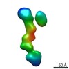
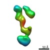
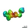
 Movie
Movie Controller

[English] 日本語
 Yorodumi
- PDB-7sar: Cryo-EM structure of TMEM106B fibrils extracted from a FTLD-TDP p... -
+ Open data

- Basic information
| Entry | Database: PDB / ID: 7sar | ||||||
|---|---|---|---|---|---|---|---|
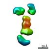
| Title | Cryo-EM structure of TMEM106B fibrils extracted from a FTLD-TDP patient, polymorph 2 | ||||||
 Components Components | Transmembrane protein 106B | ||||||
 Keywords Keywords | PROTEIN FIBRIL / TMEM106B / FTLD-TDP / amyloid | ||||||
| Function / homology |  Function and homology information Function and homology informationlysosomal protein catabolic process / regulation of lysosome organization / lysosomal lumen acidification / lysosome localization / positive regulation of dendrite development / dendrite morphogenesis / lysosomal transport / lysosome organization / neuron cellular homeostasis / late endosome membrane ...lysosomal protein catabolic process / regulation of lysosome organization / lysosomal lumen acidification / lysosome localization / positive regulation of dendrite development / dendrite morphogenesis / lysosomal transport / lysosome organization / neuron cellular homeostasis / late endosome membrane / ATPase binding / lysosome / endosome / lysosomal membrane / plasma membrane Similarity search - Function | ||||||
| Biological species |  Homo sapiens (human) Homo sapiens (human) | ||||||
| Method | ELECTRON MICROSCOPY / helical reconstruction / cryo EM / Resolution: 3.2 Å | ||||||
 Authors Authors | Cao, Q. / Jiang, Y. / Sawaya, M.R. / Eisenberg, D.S. | ||||||
| Funding support |  United States, 1items United States, 1items
| ||||||
 Citation Citation | Journal: Nature / Year: 2022 Title: Amyloid fibrils in FTLD-TDP are composed of TMEM106B and not TDP-43. Authors: Yi Xiao Jiang / Qin Cao / Michael R Sawaya / Romany Abskharon / Peng Ge / Michael DeTure / Dennis W Dickson / Janine Y Fu / Rachel R Ogorzalek Loo / Joseph A Loo / David S Eisenberg /  Abstract: Frontotemporal lobar degeneration (FTLD) is the third most common neurodegenerative condition after Alzheimer's and Parkinson's diseases. FTLD typically presents in 45 to 64 year olds with ...Frontotemporal lobar degeneration (FTLD) is the third most common neurodegenerative condition after Alzheimer's and Parkinson's diseases. FTLD typically presents in 45 to 64 year olds with behavioural changes or progressive decline of language skills. The subtype FTLD-TDP is characterized by certain clinical symptoms and pathological neuronal inclusions with TAR DNA-binding protein (TDP-43) immunoreactivity. Here we extracted amyloid fibrils from brains of four patients representing four of the five FTLD-TDP subclasses, and determined their structures by cryo-electron microscopy. Unexpectedly, all amyloid fibrils examined were composed of a 135-residue carboxy-terminal fragment of transmembrane protein 106B (TMEM106B), a lysosomal membrane protein previously implicated as a genetic risk factor for FTLD-TDP. In addition to TMEM106B fibrils, we detected abundant non-fibrillar aggregated TDP-43 by immunogold labelling. Our observations confirm that FTLD-TDP is associated with amyloid fibrils, and that the fibrils are formed by TMEM106B rather than TDP-43. | ||||||
| History |
|
- Structure visualization
| Movie |
Movie viewer |
|---|---|
| Structure viewer | Molecule: MolmilJmol/JSmol |
- Downloads & links
-Download
| PDBx/mmCIF format | 7sar.cif.gz | 262 KB | Display | PDBx/mmCIF format |
|---|---|---|---|---|
| PDB format | pdb7sar.ent.gz | 219.4 KB | Display | PDB format |
| PDBx/mmJSON format | 7sar.json.gz | Tree view | PDBx/mmJSON format | |
| Others |  Other downloads Other downloads |
-Validation report
| Summary document | 7sar_validation.pdf.gz | 795.8 KB | Display | wwPDB validaton report |
|---|---|---|---|---|
| Full document | 7sar_full_validation.pdf.gz | 825.7 KB | Display | |
| Data in XML | 7sar_validation.xml.gz | 50.9 KB | Display | |
| Data in CIF | 7sar_validation.cif.gz | 63.2 KB | Display | |
| Arichive directory | https://data.pdbj.org/pub/pdb/validation_reports/sa/7sarftp://data.pdbj.org/pub/pdb/validation_reports/sa/7sar | HTTPS FTP |
-Related structure data
| Related structure data | 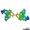 24954MC  7saqC  7sasC M: map data used to model this data C: citing same article ( |
|---|---|
| Similar structure data | |
| EM raw data | EMPIAR-11041 (Title: Cryo electron-microscopy of TMEM106B fibrils extracted from four FTLD-TDP patient brains Data size: 3.4 TB Data #1: Unaligned multi-frame micrographs of TMEM106B amyloid fibrils extracted from FTLD-TDP donor 1 [micrographs - multiframe] Data #2: Unaligned multi-frame micrographs of TMEM106B amyloid fibrils extracted from FTLD-TDP donor 2 [micrographs - multiframe] Data #3: Unaligned multi-frame micrographs of TMEM106B amyloid fibrils extracted from FTLD-TDP donor 3 [micrographs - multiframe] Data #4: Unaligned multi-frame micrographs of TMEM106B amyloid fibrils extracted from FTLD-TDP donor 4 [micrographs - multiframe]) |


-Links
 PDBj
PDBj
- Assembly
| Deposited unit | 
|
|---|---|
| 1 |
|
-Components
| #1: Protein | Mass: 31156.318 Da / Num. of mol.: 10 / Source method: isolated from a natural source Details: Glycosylation of Asn145, Asn151, Asn164, and Asn183 Source: (natural) Homo sapiens (human) / References: UniProt: Q9NUM4#2: Sugar | ChemComp-NAG /   Type: D-saccharide, beta linking / Mass: 221.208 Da / Num. of mol.: 40 / Source method: obtained synthetically / Formula: C8H15NO6 Type: D-saccharide, beta linking / Mass: 221.208 Da / Num. of mol.: 40 / Source method: obtained synthetically / Formula: C8H15NO6Has ligand of interest | N | |
|---|
-Experimental details
-Experiment
| Experiment | Method: ELECTRON MICROSCOPY |
|---|---|
| EM experiment | Aggregation state: FILAMENT / 3D reconstruction method: helical reconstruction |
- Sample preparation
| Component | Name: TMEM106B fibrils, polymorph 2 / Type: ORGANELLE OR CELLULAR COMPONENT Details: Amyloid fibrils extracted from autopsied brain tissues of a donor with FTLD-TDP Entity ID: #1 / Source: NATURAL |
|---|---|
| Molecular weight | Units: KILODALTONS/NANOMETER / Experimental value: NO |
| Source (natural) | Organism: Homo sapiens (human) |
| Buffer solution | pH: 7.5 |
| Specimen | Embedding applied: NO / Shadowing applied: NO / Staining applied: NO / Vitrification applied: YES |
| Vitrification | Instrument: FEI VITROBOT MARK IV / Cryogen name: ETHANE / Humidity: 100 % / Chamber temperature: 298 K |
- Electron microscopy imaging
| Experimental equipment |  Model: Titan Krios / Image courtesy: FEI Company |
|---|---|
| Microscopy | Model: FEI TITAN KRIOS |
| Electron gun | Electron source:  FIELD EMISSION GUN / Accelerating voltage: 300 kV / Illumination mode: FLOOD BEAM FIELD EMISSION GUN / Accelerating voltage: 300 kV / Illumination mode: FLOOD BEAM |
| Electron lens | Mode: BRIGHT FIELD |
| Image recording | Electron dose: 39 e/Å2 / Detector mode: SUPER-RESOLUTION / Film or detector model: GATAN K3 (6k x 4k) / Num. of grids imaged: 1 |
- Processing
| EM software |
| ||||||||||||||||||||||||||||||||
|---|---|---|---|---|---|---|---|---|---|---|---|---|---|---|---|---|---|---|---|---|---|---|---|---|---|---|---|---|---|---|---|---|---|
| CTF correction | Type: PHASE FLIPPING AND AMPLITUDE CORRECTION | ||||||||||||||||||||||||||||||||
| Helical symmerty | Angular rotation/subunit: 179.59 ° / Axial rise/subunit: 4.91 Å / Axial symmetry: C2 | ||||||||||||||||||||||||||||||||
| 3D reconstruction | Resolution: 3.2 Å / Resolution method: FSC 0.143 CUT-OFF / Num. of particles: 16883 / Symmetry type: HELICAL | ||||||||||||||||||||||||||||||||
| Atomic model building | B value: 93 / Protocol: FLEXIBLE FIT / Space: REAL |
