 Movie
Movie Controller


+ Open data

- Basic information
| Entry | Database: PDB / ID: 7qqk | ||||||||||||
|---|---|---|---|---|---|---|---|---|---|---|---|---|---|
| Title | TIR-SAVED effector bound to cA3 | ||||||||||||
 Components Components |
| ||||||||||||
 Keywords Keywords | SIGNALING PROTEIN / Microbacterium ketosireducens TIR SAVED complex bound to cA3 | ||||||||||||
| Function / homology |  Function and homology information Function and homology informationnucleobase-containing small molecule biosynthetic process / NAD catabolic process / NAD+ nucleosidase activity / signal transduction Similarity search - Function | ||||||||||||
| Biological species |  Microbacterium ketosireducens (bacteria) Microbacterium ketosireducens (bacteria) | ||||||||||||
| Method | ELECTRON MICROSCOPY / single particle reconstruction / cryo EM / Resolution: 3.8 Å | ||||||||||||
 Authors Authors | Spagnolo, L. / White, M.F. / Hogrel, G. / Guild, A. | ||||||||||||
| Funding support |  United Kingdom, 3items United Kingdom, 3items
| ||||||||||||
 Citation Citation | Journal: Nature / Year: 2022 Title: Cyclic nucleotide-induced helical structure activates a TIR immune effector. Authors: Gaëlle Hogrel / Abbie Guild / Shirley Graham / Hannah Rickman / Sabine Grüschow / Quentin Bertrand / Laura Spagnolo / Malcolm F White /   Abstract: Cyclic nucleotide signalling is a key component of antiviral defence in all domains of life. Viral detection activates a nucleotide cyclase to generate a second messenger, resulting in activation of ...Cyclic nucleotide signalling is a key component of antiviral defence in all domains of life. Viral detection activates a nucleotide cyclase to generate a second messenger, resulting in activation of effector proteins. This is exemplified by the metazoan cGAS-STING innate immunity pathway, which originated in bacteria. These defence systems require a sensor domain to bind the cyclic nucleotide and are often coupled with an effector domain that, when activated, causes cell death by destroying essential biomolecules. One example is the Toll/interleukin-1 receptor (TIR) domain, which degrades the essential cofactor NAD when activated in response to infection in plants and bacteria or during programmed nerve cell death. Here we show that a bacterial antiviral defence system generates a cyclic tri-adenylate that binds to a TIR-SAVED effector, acting as the 'glue' to allow assembly of an extended superhelical solenoid structure. Adjacent TIR subunits interact to organize and complete a composite active site, allowing NAD degradation. Activation requires extended filament formation, both in vitro and in vivo. Our study highlights an example of large-scale molecular assembly controlled by cyclic nucleotides and reveals key details of the mechanism of TIR enzyme activation. | ||||||||||||
| History |
|
- Structure visualization
| Structure viewer | Molecule: MolmilJmol/JSmol |
|---|
- Downloads & links
-Download
| PDBx/mmCIF format | 7qqk.cif.gz | 293.2 KB | Display | PDBx/mmCIF format |
|---|---|---|---|---|
| PDB format | pdb7qqk.ent.gz | 241.5 KB | Display | PDB format |
| PDBx/mmJSON format | 7qqk.json.gz | Tree view | PDBx/mmJSON format | |
| Others |  Other downloads Other downloads |
-Validation report
| Summary document | 7qqk_validation.pdf.gz | 1.5 MB | Display | wwPDB validaton report |
|---|---|---|---|---|
| Full document | 7qqk_full_validation.pdf.gz | 1.6 MB | Display | |
| Data in XML | 7qqk_validation.xml.gz | 70.8 KB | Display | |
| Data in CIF | 7qqk_validation.cif.gz | 103.8 KB | Display | |
| Arichive directory | https://data.pdbj.org/pub/pdb/validation_reports/qq/7qqkftp://data.pdbj.org/pub/pdb/validation_reports/qq/7qqk | HTTPS FTP |
-Related structure data
| Related structure data |  14122MC M: map data used to model this data C: citing same article ( |
|---|---|
| Similar structure data |
-Links
 PDBj
PDBj
- Assembly
| Deposited unit | 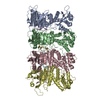
|
|---|---|
| 1 |
|
-Components
| #1: Protein | Mass: 46815.875 Da / Num. of mol.: 4 Source method: isolated from a genetically manipulated source Source: (gene. exp.) Microbacterium ketosireducens (bacteria)Gene: RS81_00402 / Production host: #2: RNA chain | Mass: 942.660 Da / Num. of mol.: 4 / Source method: obtained synthetically / Source: (synth.) Microbacterium ketosireducens (bacteria) |
|---|
-Experimental details
-Experiment
| Experiment | Method: ELECTRON MICROSCOPY |
|---|---|
| EM experiment | Aggregation state: PARTICLE / 3D reconstruction method: single particle reconstruction |
- Sample preparation
| Component | Name: one tier of TIR_SAVED bound to cA3 / Type: COMPLEX / Entity ID: all / Source: RECOMBINANT |
|---|---|
| Molecular weight | Experimental value: NO |
| Source (natural) | Organism: Microbacterium ketosireducens (bacteria) |
| Source (recombinant) | Organism: |
| Buffer solution | pH: 7.5 |
| Specimen | Embedding applied: NO / Shadowing applied: NO / Staining applied: NO / Vitrification applied: YES |
| Vitrification | Cryogen name: ETHANE |
- Electron microscopy imaging
| Microscopy | Model: JEOL CRYO ARM 300 |
|---|---|
| Electron gun | Electron source:  FIELD EMISSION GUN / Accelerating voltage: 300 kV / Illumination mode: FLOOD BEAM FIELD EMISSION GUN / Accelerating voltage: 300 kV / Illumination mode: FLOOD BEAM |
| Electron lens | Mode: BRIGHT FIELD / Nominal defocus max: 5000 nm / Nominal defocus min: 1500 nm |
| Image recording | Electron dose: 46 e/Å2 / Film or detector model: DIRECT ELECTRON DE-64 (8k x 8k) |
- Processing
| Software | Name: PHENIX / Version: 1.19.2_4158: / Classification: refinement | ||||||||||||||||||||||||
|---|---|---|---|---|---|---|---|---|---|---|---|---|---|---|---|---|---|---|---|---|---|---|---|---|---|
| CTF correction | Type: PHASE FLIPPING AND AMPLITUDE CORRECTION | ||||||||||||||||||||||||
| 3D reconstruction | Resolution: 3.8 Å / Resolution method: FSC 0.143 CUT-OFF / Num. of particles: 596378 / Symmetry type: POINT | ||||||||||||||||||||||||
| Refine LS restraints |
|
