[English] 日本語
 Yorodumi
- PDB-7qne: Cryo-EM structure of human full-length synaptic alpha1beta3gamma2... -
+ Open data
- Basic information
| Entry | Database: PDB / ID: 7qne | ||||||||||||||||||
|---|---|---|---|---|---|---|---|---|---|---|---|---|---|---|---|---|---|---|---|
| Title | Cryo-EM structure of human full-length synaptic alpha1beta3gamma2 GABA(A)R in complex with Ro15-4513 and megabody Mb38 | ||||||||||||||||||
 Components Components |
| ||||||||||||||||||
 Keywords Keywords | MEMBRANE PROTEIN / pentameric ligand-gated ion channel / neurotransmitter receptor / GABA receptor | ||||||||||||||||||
| Function / homology |  Function and homology information Function and homology informationcircadian sleep/wake cycle, REM sleep / reproductive behavior / hard palate development / cellular response to histamine / GABA receptor activation / inner ear receptor cell development / inhibitory synapse assembly / GABA-A receptor activity / GABA-gated chloride ion channel activity / GABA-A receptor complex ...circadian sleep/wake cycle, REM sleep / reproductive behavior / hard palate development / cellular response to histamine / GABA receptor activation / inner ear receptor cell development / inhibitory synapse assembly / GABA-A receptor activity / GABA-gated chloride ion channel activity / GABA-A receptor complex / innervation / response to anesthetic / postsynaptic specialization membrane / inhibitory postsynaptic potential / gamma-aminobutyric acid signaling pathway / synaptic transmission, GABAergic / cellular response to zinc ion / chloride channel activity / exploration behavior / motor behavior / roof of mouth development / Signaling by ERBB4 / cochlea development / social behavior / chloride channel complex / extracellular ligand-gated monoatomic ion channel activity / chloride transmembrane transport / cytoplasmic vesicle membrane / cerebellum development / learning / transmitter-gated monoatomic ion channel activity involved in regulation of postsynaptic membrane potential / GABA-ergic synapse / memory / dendritic spine / postsynaptic membrane / response to xenobiotic stimulus / cell surface / signal transduction / identical protein binding / plasma membrane Similarity search - Function | ||||||||||||||||||
| Biological species |  Homo sapiens (human) Homo sapiens (human) | ||||||||||||||||||
| Method | ELECTRON MICROSCOPY / single particle reconstruction / cryo EM / Resolution: 2.7 Å | ||||||||||||||||||
 Authors Authors | Sente, A. / Desai, R. / Naydenova, K. / Malinauskas, T. / Jounaidi, Y. / Miehling, J. / Zhou, X. / Masiulis, S. / Hardwick, S.W. / Chirgadze, D.Y. ...Sente, A. / Desai, R. / Naydenova, K. / Malinauskas, T. / Jounaidi, Y. / Miehling, J. / Zhou, X. / Masiulis, S. / Hardwick, S.W. / Chirgadze, D.Y. / Miller, K.W. / Aricescu, A.R. | ||||||||||||||||||
| Funding support |  United Kingdom, 5items United Kingdom, 5items
| ||||||||||||||||||
 Citation Citation |  Journal: Nature / Year: 2022 Journal: Nature / Year: 2022Title: Differential assembly diversifies GABA receptor structures and signalling. Authors: Andrija Sente / Rooma Desai / Katerina Naydenova / Tomas Malinauskas / Youssef Jounaidi / Jonas Miehling / Xiaojuan Zhou / Simonas Masiulis / Steven W Hardwick / Dimitri Y Chirgadze / Keith ...Authors: Andrija Sente / Rooma Desai / Katerina Naydenova / Tomas Malinauskas / Youssef Jounaidi / Jonas Miehling / Xiaojuan Zhou / Simonas Masiulis / Steven W Hardwick / Dimitri Y Chirgadze / Keith W Miller / A Radu Aricescu /    Abstract: Type A γ-aminobutyric acid receptors (GABARs) are pentameric ligand-gated chloride channels that mediate fast inhibitory signalling in neural circuits and can be modulated by essential medicines ...Type A γ-aminobutyric acid receptors (GABARs) are pentameric ligand-gated chloride channels that mediate fast inhibitory signalling in neural circuits and can be modulated by essential medicines including general anaesthetics and benzodiazepines. Human GABAR subunits are encoded by 19 paralogous genes that can, in theory, give rise to 495,235 receptor types. However, the principles that govern the formation of pentamers, the permutational landscape of receptors that may emerge from a subunit set and the effect that this has on GABAergic signalling remain largely unknown. Here we use cryogenic electron microscopy to determine the structures of extrasynaptic GABARs assembled from α4, β3 and δ subunits, and their counterparts incorporating γ2 instead of δ subunits. In each case, we identified two receptor subtypes with distinct stoichiometries and arrangements, all four differing from those previously observed for synaptic, α1-containing receptors. This, in turn, affects receptor responses to physiological and synthetic modulators by creating or eliminating ligand-binding sites at subunit interfaces. We provide structural and functional evidence that selected GABAR arrangements can act as coincidence detectors, simultaneously responding to two neurotransmitters: GABA and histamine. Using assembly simulations and single-cell RNA sequencing data, we calculated the upper bounds for receptor diversity in recombinant systems and in vivo. We propose that differential assembly is a pervasive mechanism for regulating the physiology and pharmacology of GABARs. | ||||||||||||||||||
| History |
|
- Structure visualization
| Structure viewer | Molecule:  Molmil Molmil Jmol/JSmol Jmol/JSmol |
|---|
- Downloads & links
- Download
| PDBx/mmCIF format |  7qne.cif.gz 7qne.cif.gz | 365.8 KB | Display |  PDBx/mmCIF format PDBx/mmCIF format |
|---|---|---|---|---|
| PDB format |  pdb7qne.ent.gz pdb7qne.ent.gz | 283.3 KB | Display |  PDB format PDB format |
| PDBx/mmJSON format |  7qne.json.gz 7qne.json.gz | Tree view |  PDBx/mmJSON format PDBx/mmJSON format | |
| Others |  Other downloads Other downloads |
-Validation report
| Arichive directory |  https://data.pdbj.org/pub/pdb/validation_reports/qn/7qne https://data.pdbj.org/pub/pdb/validation_reports/qn/7qne ftp://data.pdbj.org/pub/pdb/validation_reports/qn/7qne ftp://data.pdbj.org/pub/pdb/validation_reports/qn/7qne | HTTPS FTP |
|---|
-Related structure data
| Related structure data |  14076MC  7qn5C  7qn6C  7qn7C  7qn8C  7qn9C  7qnaC  7qnbC  7qncC  7qndC M: map data used to model this data C: citing same article ( |
|---|---|
| Similar structure data | Similarity search - Function & homology  F&H Search F&H Search |
| EM raw data |  EMPIAR-10912 (Title: Cryo-EM micrographs of GABA(A)Rs purified from cells expressing human full-length alpha1, beta3 and gamma subunits, in presence of Ro15-4513 and megabody Mb38 EMPIAR-10912 (Title: Cryo-EM micrographs of GABA(A)Rs purified from cells expressing human full-length alpha1, beta3 and gamma subunits, in presence of Ro15-4513 and megabody Mb38Data size: 1.4 TB Data #1: Unaligned multi-frame micrographs of GABA(A)Rs purified from cells expressing human full-length alpha1, beta3 and gamma subunits, in presence of Ro15-4513 and megabody Mb38 [micrographs - multiframe]) |
- Links
Links
- Assembly
| Deposited unit | 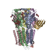
|
|---|---|
| 1 |
|
- Components
-GABA(A) receptor subunit ... , 2 types, 3 molecules ADC
| #1: Protein | Mass: 51865.664 Da / Num. of mol.: 2 Source method: isolated from a genetically manipulated source Source: (gene. exp.)  Homo sapiens (human) / Gene: GABRA1 / Cell line (production host): HEK293S TetR / Production host: Homo sapiens (human) / Gene: GABRA1 / Cell line (production host): HEK293S TetR / Production host:  Homo sapiens (human) / References: UniProt: A0A1B0GV38 Homo sapiens (human) / References: UniProt: A0A1B0GV38#3: Protein | | Mass: 56922.055 Da / Num. of mol.: 1 Source method: isolated from a genetically manipulated source Source: (gene. exp.)  Homo sapiens (human) / Gene: GABRG2 / Cell line (production host): HEK293S TetR / Production host: Homo sapiens (human) / Gene: GABRG2 / Cell line (production host): HEK293S TetR / Production host:  Homo sapiens (human) / References: UniProt: A0A286YFI6 Homo sapiens (human) / References: UniProt: A0A286YFI6 |
|---|
-Protein / Antibody , 2 types, 3 molecules BEG
| #2: Protein | Mass: 54180.348 Da / Num. of mol.: 2 Source method: isolated from a genetically manipulated source Source: (gene. exp.)  Homo sapiens (human) / Gene: GABRB3 / Cell line (production host): HEK293S TetR / Production host: Homo sapiens (human) / Gene: GABRB3 / Cell line (production host): HEK293S TetR / Production host:  Homo sapiens (human) / References: UniProt: P28472 Homo sapiens (human) / References: UniProt: P28472#4: Antibody | | Mass: 13462.909 Da / Num. of mol.: 1 Source method: isolated from a genetically manipulated source Source: (gene. exp.)   |
|---|
-Sugars , 5 types, 7 molecules 
| #5: Polysaccharide | alpha-D-mannopyranose-(1-2)-alpha-D-mannopyranose-(1-2)-alpha-D-mannopyranose-(1-3)-[alpha-D- ...alpha-D-mannopyranose-(1-2)-alpha-D-mannopyranose-(1-2)-alpha-D-mannopyranose-(1-3)-[alpha-D-mannopyranose-(1-3)-[alpha-D-mannopyranose-(1-6)]alpha-D-mannopyranose-(1-6)]beta-D-mannopyranose-(1-4)-2-acetamido-2-deoxy-beta-D-glucopyranose-(1-4)-2-acetamido-2-deoxy-beta-D-glucopyranose Source method: isolated from a genetically manipulated source | ||||||
|---|---|---|---|---|---|---|---|
| #6: Polysaccharide | Source method: isolated from a genetically manipulated source #7: Polysaccharide | Source method: isolated from a genetically manipulated source #8: Polysaccharide | alpha-D-mannopyranose-(1-3)-[alpha-D-mannopyranose-(1-6)]beta-D-mannopyranose-(1-4)-2-acetamido-2- ...alpha-D-mannopyranose-(1-3)-[alpha-D-mannopyranose-(1-6)]beta-D-mannopyranose-(1-4)-2-acetamido-2-deoxy-beta-D-glucopyranose-(1-4)-2-acetamido-2-deoxy-beta-D-glucopyranose | Source method: isolated from a genetically manipulated source #13: Sugar | ChemComp-NAG / | |
-Non-polymers , 5 types, 11 molecules 



| #9: Chemical | | #10: Chemical | ChemComp-OCT / | #11: Chemical | ChemComp-D10 / #12: Chemical | #14: Chemical | ChemComp-EIE / | |
|---|
-Details
| Has ligand of interest | Y |
|---|---|
| Has protein modification | Y |
-Experimental details
-Experiment
| Experiment | Method: ELECTRON MICROSCOPY |
|---|---|
| EM experiment | Aggregation state: PARTICLE / 3D reconstruction method: single particle reconstruction |
- Sample preparation
| Component | Name: Human full-length synaptic alpha1beta3gamma2 GABA(A)R in complex with Ro15-4513 and megabody Mb38 Type: COMPLEX / Entity ID: #1-#4 / Source: RECOMBINANT | |||||||||||||||
|---|---|---|---|---|---|---|---|---|---|---|---|---|---|---|---|---|
| Source (natural) | Organism:  Homo sapiens (human) Homo sapiens (human) | |||||||||||||||
| Source (recombinant) | Organism:  Homo sapiens (human) / Cell: HEK293S TetR Homo sapiens (human) / Cell: HEK293S TetR | |||||||||||||||
| Buffer solution | pH: 7.6 | |||||||||||||||
| Buffer component |
| |||||||||||||||
| Specimen | Embedding applied: NO / Shadowing applied: NO / Staining applied: NO / Vitrification applied: YES | |||||||||||||||
| Vitrification | Instrument: LEICA PLUNGER / Cryogen name: ETHANE / Humidity: 95 % / Chamber temperature: 287 K |
- Electron microscopy imaging
| Experimental equipment |  Model: Titan Krios / Image courtesy: FEI Company |
|---|---|
| Microscopy | Model: FEI TITAN KRIOS |
| Electron gun | Electron source:  FIELD EMISSION GUN / Accelerating voltage: 300 kV / Illumination mode: FLOOD BEAM FIELD EMISSION GUN / Accelerating voltage: 300 kV / Illumination mode: FLOOD BEAM |
| Electron lens | Mode: BRIGHT FIELD / Nominal magnification: 130000 X / Nominal defocus max: 2100 nm / Nominal defocus min: 700 nm / Cs: 2.7 mm / C2 aperture diameter: 50 µm / Alignment procedure: COMA FREE |
| Specimen holder | Cryogen: NITROGEN |
| Image recording | Electron dose: 40 e/Å2 / Film or detector model: GATAN K3 (6k x 4k) |
| EM imaging optics | Energyfilter name: GIF Bioquantum / Energyfilter slit width: 20 eV |
- Processing
| Software | Name: PHENIX / Version: 1.19.2_4158: / Classification: refinement | ||||||||||||||||||||||||||||||||||||||||
|---|---|---|---|---|---|---|---|---|---|---|---|---|---|---|---|---|---|---|---|---|---|---|---|---|---|---|---|---|---|---|---|---|---|---|---|---|---|---|---|---|---|
| EM software |
| ||||||||||||||||||||||||||||||||||||||||
| CTF correction | Type: PHASE FLIPPING AND AMPLITUDE CORRECTION | ||||||||||||||||||||||||||||||||||||||||
| 3D reconstruction | Resolution: 2.7 Å / Resolution method: FSC 0.143 CUT-OFF / Num. of particles: 119901 / Symmetry type: POINT | ||||||||||||||||||||||||||||||||||||||||
| Atomic model building | Protocol: AB INITIO MODEL | ||||||||||||||||||||||||||||||||||||||||
| Refine LS restraints |
|
 Movie
Movie Controller


 PDBj
PDBj


