 Movie
Movie Controller


+ Open data

- Basic information
| Entry | Database: PDB / ID: 7oae | ||||||
|---|---|---|---|---|---|---|---|
| Title | Cryo-EM structure of the plectasin fibril (double strands) | ||||||
 Components Components | Fungal defensin plectasin | ||||||
 Keywords Keywords | ANTIMICROBIAL PROTEIN / fibril / helical | ||||||
| Function / homology |  Function and homology information Function and homology informationpotassium channel regulator activity / toxin activity / defense response to bacterium / host cell plasma membrane / extracellular region / membrane Similarity search - Function | ||||||
| Biological species |  Pseudoplectania nigrella (fungus) Pseudoplectania nigrella (fungus) | ||||||
| Method | ELECTRON MICROSCOPY / helical reconstruction / cryo EM / Resolution: 2 Å | ||||||
 Authors Authors | Effantin, G. | ||||||
| Funding support | European Union, 1items
| ||||||
 Citation Citation | Journal: Nat Commun / Year: 2022 Title: pH- and concentration-dependent supramolecular assembly of a fungal defensin plectasin variant into helical non-amyloid fibrils. Authors: Christin Pohl / Gregory Effantin / Eaazhisai Kandiah / Sebastian Meier / Guanghong Zeng / Werner Streicher / Dorotea Raventos Segura / Per H Mygind / Dorthe Sandvang / Line Anker Nielsen / ...Authors: Christin Pohl / Gregory Effantin / Eaazhisai Kandiah / Sebastian Meier / Guanghong Zeng / Werner Streicher / Dorotea Raventos Segura / Per H Mygind / Dorthe Sandvang / Line Anker Nielsen / Günther H J Peters / Guy Schoehn / Christoph Mueller-Dieckmann / Allan Noergaard / Pernille Harris /     Abstract: Self-assembly and fibril formation play important roles in protein behaviour. Amyloid fibril formation is well-studied due to its role in neurodegenerative diseases and characterized by refolding of ...Self-assembly and fibril formation play important roles in protein behaviour. Amyloid fibril formation is well-studied due to its role in neurodegenerative diseases and characterized by refolding of the protein into predominantly β-sheet form. However, much less is known about the assembly of proteins into other types of supramolecular structures. Using cryo-electron microscopy at a resolution of 1.97 Å, we show that a triple-mutant of the anti-microbial peptide plectasin, PPI42, assembles into helical non-amyloid fibrils. The in vitro anti-microbial activity was determined and shown to be enhanced compared to the wildtype. Plectasin contains a cysteine-stabilised α-helix-β-sheet structure, which remains intact upon fibril formation. Two protofilaments form a right-handed protein fibril. The fibril formation is reversible and follows sigmoidal kinetics with a pH- and concentration dependent equilibrium between soluble monomer and protein fibril. This high-resolution structure reveals that α/β proteins can natively assemble into fibrils. | ||||||
| History |
|
- Structure visualization
| Structure viewer | Molecule: MolmilJmol/JSmol |
|---|
- Downloads & links
-Download
| PDBx/mmCIF format | 7oae.cif.gz | 275.4 KB | Display | PDBx/mmCIF format |
|---|---|---|---|---|
| PDB format | pdb7oae.ent.gz | 235.2 KB | Display | PDB format |
| PDBx/mmJSON format | 7oae.json.gz | Tree view | PDBx/mmJSON format | |
| Others |  Other downloads Other downloads |
-Validation report
| Summary document | 7oae_validation.pdf.gz | 862.5 KB | Display | wwPDB validaton report |
|---|---|---|---|---|
| Full document | 7oae_full_validation.pdf.gz | 861.7 KB | Display | |
| Data in XML | 7oae_validation.xml.gz | 43.9 KB | Display | |
| Data in CIF | 7oae_validation.cif.gz | 66.3 KB | Display | |
| Arichive directory | https://data.pdbj.org/pub/pdb/validation_reports/oa/7oaeftp://data.pdbj.org/pub/pdb/validation_reports/oa/7oae | HTTPS FTP |
-Related structure data
| Related structure data | 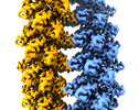 12775MC  7o76C  7oagC M: map data used to model this data C: citing same article ( |
|---|---|
| Similar structure data |

-Links
 PDBj
PDBj

- Assembly
| Deposited unit | 
|
|---|---|
| 1 |
|
-Components
| #1: Protein/peptide | Mass: 4402.092 Da / Num. of mol.: 42 Source method: isolated from a genetically manipulated source Source: (gene. exp.) Pseudoplectania nigrella (fungus) / Gene: DEF / Production host:  |
|---|
-Experimental details
-Experiment
| Experiment | Method: ELECTRON MICROSCOPY |
|---|---|
| EM experiment | Aggregation state: HELICAL ARRAY / 3D reconstruction method: helical reconstruction |
- Sample preparation
| Component | Name: supermolecular assembly of plectasin into two protein fibrils Type: COMPLEX / Entity ID: all / Source: RECOMBINANT |
|---|---|
| Molecular weight | Experimental value: NO |
| Source (natural) | Organism: Pseudoplectania nigrella (fungus) |
| Source (recombinant) | Organism: |
| Buffer solution | pH: 5.5 |
| Specimen | Embedding applied: NO / Shadowing applied: NO / Staining applied: NO / Vitrification applied: YES |
| Vitrification | Cryogen name: ETHANE |
- Electron microscopy imaging
| Experimental equipment |  Model: Titan Krios / Image courtesy: FEI Company |
|---|---|
| Microscopy | Model: FEI TITAN KRIOS |
| Electron gun | Electron source:  FIELD EMISSION GUN / Accelerating voltage: 300 kV / Illumination mode: FLOOD BEAM FIELD EMISSION GUN / Accelerating voltage: 300 kV / Illumination mode: FLOOD BEAM |
| Electron lens | Mode: BRIGHT FIELD |
| Image recording | Electron dose: 40 e/Å2 / Film or detector model: GATAN K2 SUMMIT (4k x 4k) |
- Processing
| Software | Name: PHENIX / Version: 1.18.2_3874: / Classification: refinement | ||||||||||||||||||||||||
|---|---|---|---|---|---|---|---|---|---|---|---|---|---|---|---|---|---|---|---|---|---|---|---|---|---|
| CTF correction | Type: PHASE FLIPPING AND AMPLITUDE CORRECTION | ||||||||||||||||||||||||
| Helical symmerty | Angular rotation/subunit: 15.75 ° / Axial rise/subunit: 25.1 Å / Axial symmetry: C2 | ||||||||||||||||||||||||
| 3D reconstruction | Resolution: 2 Å / Resolution method: FSC 0.143 CUT-OFF / Num. of particles: 764822 / Symmetry type: HELICAL | ||||||||||||||||||||||||
| Refine LS restraints |
|
