 ムービー
ムービー コントローラー


+ データを開く

- 基本情報
基本情報
| 登録情報 | データベース: PDB / ID: 7k5b | ||||||
|---|---|---|---|---|---|---|---|
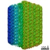
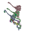
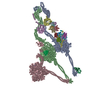
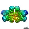
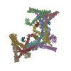
| タイトル | Structure of outer-arm dynein bound to microtubule doublet in microtubule binding state 2 (MTBS-2) | ||||||
 要素 要素 |
| ||||||
 キーワード キーワード | MOTOR PROTEIN / three head / outer dynein arms / microtubule binding | ||||||
| 機能・相同性 |  機能・相同性情報 機能・相同性情報inner dynein arm / axonemal dynein complex / outer dynein arm / outer dynein arm assembly / inner dynein arm assembly / cilium movement involved in cell motility / cilium movement / dynein light chain binding / dynein heavy chain binding / dynein complex ...inner dynein arm / axonemal dynein complex / outer dynein arm / outer dynein arm assembly / inner dynein arm assembly / cilium movement involved in cell motility / cilium movement / dynein light chain binding / dynein heavy chain binding / dynein complex / myosin II complex / minus-end-directed microtubule motor activity / dynein light intermediate chain binding / cytoplasmic dynein complex / microtubule-based movement / dynein intermediate chain binding / axoneme / mRNA transport / microtubule-based process / protein transport / microtubule binding / microtubule / calcium ion binding / ATP binding / nucleus / cytoplasm 類似検索 - 分子機能 | ||||||
| 生物種 |   Tetrahymena thermophila (真核生物) Tetrahymena thermophila (真核生物) | ||||||
| 手法 | 電子顕微鏡法 / 単粒子再構成法 / クライオ電子顕微鏡法 / 解像度: 4.5 Å | ||||||
 データ登録者 データ登録者 | Rao, Q. / Zhang, K. | ||||||
 引用 引用 | ジャーナル: Nat Struct Mol Biol / 年: 2021 タイトル: Structures of outer-arm dynein array on microtubule doublet reveal a motor coordination mechanism. 著者: Qinhui Rao / Long Han / Yue Wang / Pengxin Chai / Yin-Wei Kuo / Renbin Yang / Fangheng Hu / Yuchen Yang / Jonathon Howard / Kai Zhang /  要旨: Thousands of outer-arm dyneins (OADs) are arrayed in the axoneme to drive a rhythmic ciliary beat. Coordination among multiple OADs is essential for generating mechanical forces to bend microtubule ...Thousands of outer-arm dyneins (OADs) are arrayed in the axoneme to drive a rhythmic ciliary beat. Coordination among multiple OADs is essential for generating mechanical forces to bend microtubule doublets (MTDs). Using electron microscopy, we determined high-resolution structures of Tetrahymena thermophila OAD arrays bound to MTDs in two different states. OAD preferentially binds to MTD protofilaments with a pattern resembling the native tracks for its distinct microtubule-binding domains. Upon MTD binding, free OADs are induced to adopt a stable parallel conformation, primed for array formation. Extensive tail-to-head (TTH) interactions between OADs are observed, which need to be broken for ATP turnover by the dynein motor. We propose that OADs in an array sequentially hydrolyze ATP to slide the MTDs. ATP hydrolysis in turn relaxes the TTH interfaces to effect free nucleotide cycles of downstream OADs. These findings lead to a model explaining how conformational changes in the axoneme produce coordinated action of dyneins. | ||||||
| 履歴 |
|
- 構造の表示
構造の表示
| ムービー |
ムービービューア |
|---|---|
| 構造ビューア | 分子: MolmilJmol/JSmol |
- ダウンロードとリンク
-ダウンロード
| PDBx/mmCIF形式 | 7k5b.cif.gz | 2.9 MB | 表示 | PDBx/mmCIF形式 |
|---|---|---|---|---|
| PDB形式 | pdb7k5b.ent.gz | 表示 | PDB形式 | |
| PDBx/mmJSON形式 | 7k5b.json.gz | ツリー表示 | PDBx/mmJSON形式 | |
| その他 |  その他のダウンロード その他のダウンロード |
-検証レポート
| アーカイブディレクトリ | https://data.pdbj.org/pub/pdb/validation_reports/k5/7k5bftp://data.pdbj.org/pub/pdb/validation_reports/k5/7k5b | HTTPS FTP |
|---|
-関連構造データ


-リンク
 PDBj
PDBj


- 集合体
集合体
| 登録構造単位 | 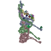
|
|---|---|
| 1 |
|
-要素
-タンパク質 , 5種, 5分子 ACDEP
| #1: タンパク質 | 分子量: 533780.188 Da / 分子数: 1 / 由来タイプ: 天然 / 由来: (天然) Tetrahymena thermophila (真核生物) / 参照: UniProt: Q22A67 |
|---|---|
| #3: タンパク質 | 分子量: 450623.219 Da / 分子数: 1 / 由来タイプ: 天然 / 由来: (天然) Tetrahymena thermophila (真核生物) |
| #4: タンパク質 | 分子量: 69514.820 Da / 分子数: 1 / 由来タイプ: 天然 / 由来: (天然) Tetrahymena thermophila (真核生物) / 参照: UniProt: I7M008 |
| #5: タンパク質 | 分子量: 63702.582 Da / 分子数: 1 / 由来タイプ: 天然 / 由来: (天然) Tetrahymena thermophila (真核生物) / 参照: UniProt: Q23FU1 |
| #16: タンパク質 | 分子量: 12987.904 Da / 分子数: 1 / 由来タイプ: 天然 / 由来: (天然) Tetrahymena thermophila (真核生物) / 参照: UniProt: A4VD75 |
-Dynein light ... , 12種, 12分子 FGHIJKLMNOQR
| #6: タンパク質 | 分子量: 14193.171 Da / 分子数: 1 / 由来タイプ: 天然 / 由来: (天然) Tetrahymena thermophila (真核生物) / 参照: UniProt: I7MHB1 |
|---|---|
| #7: タンパク質 | 分子量: 17470.111 Da / 分子数: 1 / 由来タイプ: 天然 / 由来: (天然) Tetrahymena thermophila (真核生物) / 参照: UniProt: Q22MV2 |
| #8: タンパク質 | 分子量: 10649.161 Da / 分子数: 1 / 由来タイプ: 天然 / 由来: (天然) Tetrahymena thermophila (真核生物) / 参照: UniProt: Q1HFW2 |
| #9: タンパク質 | 分子量: 11830.534 Da / 分子数: 1 / 由来タイプ: 天然 / 由来: (天然) Tetrahymena thermophila (真核生物) / 参照: UniProt: Q1HFX0 |
| #10: タンパク質 | 分子量: 11435.072 Da / 分子数: 1 / 由来タイプ: 天然 / 由来: (天然) Tetrahymena thermophila (真核生物) / 参照: UniProt: Q22R86 |
| #11: タンパク質 | 分子量: 10670.071 Da / 分子数: 1 / 由来タイプ: 天然 / 由来: (天然) Tetrahymena thermophila (真核生物) / 参照: UniProt: Q1HFV9 |
| #12: タンパク質 | 分子量: 12516.457 Da / 分子数: 1 / 由来タイプ: 天然 / 由来: (天然) Tetrahymena thermophila (真核生物) / 参照: UniProt: W7XJB1 |
| #13: タンパク質 | 分子量: 10453.167 Da / 分子数: 1 / 由来タイプ: 天然 / 由来: (天然) Tetrahymena thermophila (真核生物) / 参照: UniProt: Q1HFW0 |
| #14: タンパク質 | 分子量: 12856.455 Da / 分子数: 1 / 由来タイプ: 天然 / 由来: (天然) Tetrahymena thermophila (真核生物) / 参照: UniProt: A4VEB3 |
| #15: タンパク質 | 分子量: 14117.313 Da / 分子数: 1 / 由来タイプ: 天然 / 由来: (天然) Tetrahymena thermophila (真核生物) / 参照: UniProt: Q1HGH8 |
| #17: タンパク質 | 分子量: 21705.334 Da / 分子数: 1 / 由来タイプ: 天然 / 由来: (天然) Tetrahymena thermophila (真核生物) / 参照: UniProt: Q1HGH9 |
| #18: タンパク質 | 分子量: 16803.703 Da / 分子数: 1 / 由来タイプ: 天然 / 由来: (天然) Tetrahymena thermophila (真核生物) / 参照: UniProt: Q1HFX4 |
-抗体 , 1種, 1分子 B
| #2: 抗体 | 分子量: 529207.188 Da / 分子数: 1 / 由来タイプ: 天然 / 由来: (天然) Tetrahymena thermophila (真核生物) / 参照: UniProt: I7M9J2 |
|---|
-非ポリマー , 3種, 18分子 


| #19: 化合物 | ChemComp-ADP /  分子量: 427.201 Da / 分子数: 6 / 由来タイプ: 合成 / 式: C10H15N5O10P2 / コメント: ADP, エネルギー貯蔵分子*YM 分子量: 427.201 Da / 分子数: 6 / 由来タイプ: 合成 / 式: C10H15N5O10P2 / コメント: ADP, エネルギー貯蔵分子*YM#20: 化合物 |  分子量: 507.181 Da / 分子数: 3 / 由来タイプ: 合成 / 式: C10H16N5O13P3 / コメント: ATP, エネルギー貯蔵分子*YM 分子量: 507.181 Da / 分子数: 3 / 由来タイプ: 合成 / 式: C10H16N5O13P3 / コメント: ATP, エネルギー貯蔵分子*YM#21: 化合物 | ChemComp-MG /  分子量: 24.305 Da / 分子数: 9 / 由来タイプ: 合成 / 式: Mg 分子量: 24.305 Da / 分子数: 9 / 由来タイプ: 合成 / 式: Mg |
|---|
-詳細
| 研究の焦点であるリガンドがあるか | N |
|---|---|
| Has protein modification | Y |
-実験情報
-実験
| 実験 | 手法: 電子顕微鏡法 |
|---|---|
| EM実験 | 試料の集合状態: FILAMENT / 3次元再構成法: 単粒子再構成法 |
- 試料調製
試料調製
| 構成要素 | 名称: Outer-arm dynein bound to microtubule doublet in microtubule binding state 2 (MTBS-2) タイプ: COMPLEX / Entity ID: #1-#18 / 由来: NATURAL |
|---|---|
| 由来(天然) | 生物種: Tetrahymena thermophila (真核生物) |
| 緩衝液 | pH: 7.4 |
| 試料 | 包埋: NO / シャドウイング: NO / 染色: NO / 凍結: YES |
| 急速凍結 | 凍結剤: ETHANE |
- 電子顕微鏡撮影
| 実験機器 |  モデル: Titan Krios / 画像提供: FEI Company |
|---|---|
| 顕微鏡 | モデル: FEI TITAN KRIOS |
| 電子銃 | 電子線源:  FIELD EMISSION GUN / 加速電圧: 300 kV / 照射モード: FLOOD BEAM FIELD EMISSION GUN / 加速電圧: 300 kV / 照射モード: FLOOD BEAM |
| 電子レンズ | モード: BRIGHT FIELD |
| 撮影 | 電子線照射量: 53.3 e/Å2 フィルム・検出器のモデル: GATAN K2 QUANTUM (4k x 4k) |
- 解析
解析
| CTF補正 | タイプ: NONE |
|---|---|
| 3次元再構成 | 解像度: 4.5 Å / 解像度の算出法: FSC 0.143 CUT-OFF / 粒子像の数: 76936 / 対称性のタイプ: POINT |
