 Movie
Movie Controller

[English] 日本語
 Yorodumi
- PDB-7k58: Structure of outer-arm dyneins bound to microtubule with microtub... -
+ Open data

- Basic information
| Entry | Database: PDB / ID: 7k58 | ||||||
|---|---|---|---|---|---|---|---|
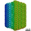
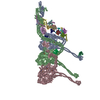
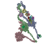
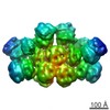
| Title | Structure of outer-arm dyneins bound to microtubule with microtubule binding state 1(MTBS-1) | ||||||
 Components Components |
| ||||||
 Keywords Keywords | STRUCTURAL PROTEIN / outer-arm dynein / axonemal dynein / microtubule doublet | ||||||
| Function / homology |  Function and homology information Function and homology informationinner dynein arm / axonemal dynein complex / outer dynein arm / outer dynein arm assembly / inner dynein arm assembly / cilium movement involved in cell motility / cilium movement / dynein light chain binding / dynein heavy chain binding / dynein complex ...inner dynein arm / axonemal dynein complex / outer dynein arm / outer dynein arm assembly / inner dynein arm assembly / cilium movement involved in cell motility / cilium movement / dynein light chain binding / dynein heavy chain binding / dynein complex / minus-end-directed microtubule motor activity / dynein light intermediate chain binding / cytoplasmic dynein complex / microtubule-based movement / dynein intermediate chain binding / axoneme / mRNA transport / microtubule-based process / protein transport / microtubule binding / microtubule / ATP binding / nucleus / cytoplasm Similarity search - Function | ||||||
| Biological species |   Tetrahymena thermophila (eukaryote) Tetrahymena thermophila (eukaryote) | ||||||
| Method | ELECTRON MICROSCOPY / single particle reconstruction / cryo EM / Resolution: 4 Å | ||||||
 Authors Authors | Qinhui, R. / Kai, Z. | ||||||
| Funding support |  United States, 1items United States, 1items
| ||||||
 Citation Citation | Journal: Nat Struct Mol Biol / Year: 2021 Title: Structures of outer-arm dynein array on microtubule doublet reveal a motor coordination mechanism. Authors: Qinhui Rao / Long Han / Yue Wang / Pengxin Chai / Yin-Wei Kuo / Renbin Yang / Fangheng Hu / Yuchen Yang / Jonathon Howard / Kai Zhang / Abstract: Thousands of outer-arm dyneins (OADs) are arrayed in the axoneme to drive a rhythmic ciliary beat. Coordination among multiple OADs is essential for generating mechanical forces to bend microtubule ...Thousands of outer-arm dyneins (OADs) are arrayed in the axoneme to drive a rhythmic ciliary beat. Coordination among multiple OADs is essential for generating mechanical forces to bend microtubule doublets (MTDs). Using electron microscopy, we determined high-resolution structures of Tetrahymena thermophila OAD arrays bound to MTDs in two different states. OAD preferentially binds to MTD protofilaments with a pattern resembling the native tracks for its distinct microtubule-binding domains. Upon MTD binding, free OADs are induced to adopt a stable parallel conformation, primed for array formation. Extensive tail-to-head (TTH) interactions between OADs are observed, which need to be broken for ATP turnover by the dynein motor. We propose that OADs in an array sequentially hydrolyze ATP to slide the MTDs. ATP hydrolysis in turn relaxes the TTH interfaces to effect free nucleotide cycles of downstream OADs. These findings lead to a model explaining how conformational changes in the axoneme produce coordinated action of dyneins. | ||||||
| History |
|
- Structure visualization
| Movie |
Movie viewer |
|---|---|
| Structure viewer | Molecule: MolmilJmol/JSmol |
- Downloads & links
-Download
| PDBx/mmCIF format | 7k58.cif.gz | 3 MB | Display | PDBx/mmCIF format |
|---|---|---|---|---|
| PDB format | pdb7k58.ent.gz | Display | PDB format | |
| PDBx/mmJSON format | 7k58.json.gz | Tree view | PDBx/mmJSON format | |
| Others |  Other downloads Other downloads |
-Validation report
| Arichive directory | https://data.pdbj.org/pub/pdb/validation_reports/k5/7k58ftp://data.pdbj.org/pub/pdb/validation_reports/k5/7k58 | HTTPS FTP |
|---|
-Related structure data
| Related structure data |  22677MC  7k5bC  7kekC  7mwgC  7n32C M: map data used to model this data C: citing same article ( |
|---|---|
| Similar structure data |


-Links
 PDBj
PDBj


- Assembly
| Deposited unit | 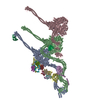
|
|---|---|
| 1 |
|
-Components
-Protein , 5 types, 5 molecules ACEDP
| #1: Protein | Mass: 533781.125 Da / Num. of mol.: 1 / Source method: isolated from a natural source / Source: (natural) Tetrahymena thermophila (eukaryote) / References: UniProt: Q22A67 |
|---|---|
| #2: Protein | Mass: 450151.656 Da / Num. of mol.: 1 / Source method: isolated from a natural source / Source: (natural) Tetrahymena thermophila (eukaryote) |
| #11: Protein | Mass: 63702.582 Da / Num. of mol.: 1 / Source method: isolated from a natural source / Source: (natural) Tetrahymena thermophila (eukaryote) / References: UniProt: Q23FU1 |
| #12: Protein | Mass: 69514.820 Da / Num. of mol.: 1 / Source method: isolated from a natural source / Source: (natural) Tetrahymena thermophila (eukaryote) / References: UniProt: I7M008 |
| #13: Protein | Mass: 12987.904 Da / Num. of mol.: 1 / Source method: isolated from a natural source / Source: (natural) Tetrahymena thermophila (eukaryote) / References: UniProt: A4VD75 |
-Dynein light ... , 11 types, 11 molecules QIHGFNOLKJM
| #3: Protein | Mass: 21737.312 Da / Num. of mol.: 1 / Source method: isolated from a natural source / Source: (natural) Tetrahymena thermophila (eukaryote) / References: UniProt: Q1HGH9 |
|---|---|
| #5: Protein | Mass: 11830.534 Da / Num. of mol.: 1 / Source method: isolated from a natural source / Source: (natural) Tetrahymena thermophila (eukaryote) / References: UniProt: Q1HFX0 |
| #6: Protein | Mass: 10649.161 Da / Num. of mol.: 1 / Source method: isolated from a natural source / Source: (natural) Tetrahymena thermophila (eukaryote) / References: UniProt: Q1HFW2 |
| #7: Protein | Mass: 10741.309 Da / Num. of mol.: 1 / Source method: isolated from a natural source / Source: (natural) Tetrahymena thermophila (eukaryote) / References: UniProt: Q1HFX1 |
| #8: Protein | Mass: 12293.036 Da / Num. of mol.: 1 / Source method: isolated from a natural source / Source: (natural) Tetrahymena thermophila (eukaryote) / References: UniProt: I7MHB1 |
| #9: Protein | Mass: 12899.480 Da / Num. of mol.: 1 / Source method: isolated from a natural source / Source: (natural) Tetrahymena thermophila (eukaryote) / References: UniProt: A4VEB3 |
| #10: Protein | Mass: 14117.313 Da / Num. of mol.: 1 / Source method: isolated from a natural source / Source: (natural) Tetrahymena thermophila (eukaryote) / References: UniProt: Q1HGH8 |
| #14: Protein | Mass: 11108.031 Da / Num. of mol.: 1 / Source method: isolated from a natural source / Source: (natural) Tetrahymena thermophila (eukaryote) / References: UniProt: W7XJB1 |
| #15: Protein | Mass: 10670.071 Da / Num. of mol.: 1 / Source method: isolated from a natural source / Source: (natural) Tetrahymena thermophila (eukaryote) / References: UniProt: Q1HFV9 |
| #16: Protein | Mass: 11403.008 Da / Num. of mol.: 1 / Source method: isolated from a natural source / Source: (natural) Tetrahymena thermophila (eukaryote) / References: UniProt: Q22R86 |
| #17: Protein | Mass: 10453.167 Da / Num. of mol.: 1 / Source method: isolated from a natural source / Source: (natural) Tetrahymena thermophila (eukaryote) / References: UniProt: Q1HFW0 |
-Antibody , 1 types, 1 molecules B
| #4: Antibody | Mass: 529322.312 Da / Num. of mol.: 1 / Source method: isolated from a natural source / Source: (natural) Tetrahymena thermophila (eukaryote) / References: UniProt: I7M9J2 |
|---|
-Non-polymers , 3 types, 18 molecules 


| #18: Chemical | ChemComp-ADP /  Mass: 427.201 Da / Num. of mol.: 6 / Source method: obtained synthetically / Formula: C10H15N5O10P2 / Comment: ADP, energy-carrying molecule*YM Mass: 427.201 Da / Num. of mol.: 6 / Source method: obtained synthetically / Formula: C10H15N5O10P2 / Comment: ADP, energy-carrying molecule*YM#19: Chemical |  Mass: 507.181 Da / Num. of mol.: 3 / Source method: obtained synthetically / Formula: C10H16N5O13P3 / Comment: ATP, energy-carrying molecule*YM Mass: 507.181 Da / Num. of mol.: 3 / Source method: obtained synthetically / Formula: C10H16N5O13P3 / Comment: ATP, energy-carrying molecule*YM#20: Chemical | ChemComp-MG /  Mass: 24.305 Da / Num. of mol.: 9 / Source method: obtained synthetically / Formula: Mg Mass: 24.305 Da / Num. of mol.: 9 / Source method: obtained synthetically / Formula: Mg |
|---|
-Details
| Has ligand of interest | N |
|---|---|
| Has protein modification | Y |
-Experimental details
-Experiment
| Experiment | Method: ELECTRON MICROSCOPY |
|---|---|
| EM experiment | Aggregation state: FILAMENT / 3D reconstruction method: single particle reconstruction |
- Sample preparation
| Component | Name: Outer-arm dynein / Type: COMPLEX / Entity ID: #1-#17 / Source: NATURAL |
|---|---|
| Molecular weight | Value: 2 MDa / Experimental value: YES |
| Source (natural) | Organism: Tetrahymena thermophila (eukaryote) |
| Buffer solution | pH: 7.4 |
| Specimen | Embedding applied: NO / Shadowing applied: NO / Staining applied: NO / Vitrification applied: YES |
| Vitrification | Instrument: FEI VITROBOT MARK IV / Cryogen name: ETHANE / Humidity: 100 % / Chamber temperature: 4 K |
- Electron microscopy imaging
| Experimental equipment |  Model: Titan Krios / Image courtesy: FEI Company |
|---|---|
| Microscopy | Model: FEI TITAN KRIOS |
| Electron gun | Electron source:  FIELD EMISSION GUN / Accelerating voltage: 300 kV / Illumination mode: FLOOD BEAM FIELD EMISSION GUN / Accelerating voltage: 300 kV / Illumination mode: FLOOD BEAM |
| Electron lens | Mode: BRIGHT FIELD |
| Specimen holder | Cryogen: NITROGEN / Specimen holder model: FEI TITAN KRIOS AUTOGRID HOLDER |
| Image recording | Electron dose: 53.3 e/Å2 / Detector mode: SUPER-RESOLUTION / Film or detector model: GATAN K2 QUANTUM (4k x 4k) |
- Processing
| Software | Name: REFMAC / Version: 5.8.0257 / Classification: refinement | ||||||||||||||||||||||||||||||||||||||||||||||||||||||||||||||||||||||||||||||||||||||||||||||||||||||||||
|---|---|---|---|---|---|---|---|---|---|---|---|---|---|---|---|---|---|---|---|---|---|---|---|---|---|---|---|---|---|---|---|---|---|---|---|---|---|---|---|---|---|---|---|---|---|---|---|---|---|---|---|---|---|---|---|---|---|---|---|---|---|---|---|---|---|---|---|---|---|---|---|---|---|---|---|---|---|---|---|---|---|---|---|---|---|---|---|---|---|---|---|---|---|---|---|---|---|---|---|---|---|---|---|---|---|---|---|
| CTF correction | Type: NONE | ||||||||||||||||||||||||||||||||||||||||||||||||||||||||||||||||||||||||||||||||||||||||||||||||||||||||||
| 3D reconstruction | Resolution: 4 Å / Resolution method: FSC 0.143 CUT-OFF / Num. of particles: 191776 / Symmetry type: POINT | ||||||||||||||||||||||||||||||||||||||||||||||||||||||||||||||||||||||||||||||||||||||||||||||||||||||||||
| Refinement | Resolution: 4→4 Å / Cor.coef. Fo:Fc: 0.925 / SU B: 15.843 / SU ML: 0.24 / ESU R: 0.288 Stereochemistry target values: MAXIMUM LIKELIHOOD WITH PHASES Details: HYDROGENS HAVE BEEN ADDED IN THE RIDING POSITIONS
| ||||||||||||||||||||||||||||||||||||||||||||||||||||||||||||||||||||||||||||||||||||||||||||||||||||||||||
| Solvent computation | Ion probe radii: 0.8 Å / Shrinkage radii: 0.8 Å / VDW probe radii: 1.2 Å / Solvent model: MASK | ||||||||||||||||||||||||||||||||||||||||||||||||||||||||||||||||||||||||||||||||||||||||||||||||||||||||||
| Displacement parameters | Biso mean: 204.36 Å2
| ||||||||||||||||||||||||||||||||||||||||||||||||||||||||||||||||||||||||||||||||||||||||||||||||||||||||||
| Refinement step | Cycle: 1 / Total: 117936 | ||||||||||||||||||||||||||||||||||||||||||||||||||||||||||||||||||||||||||||||||||||||||||||||||||||||||||
| Refine LS restraints |
|