+ Open data
- Basic information
| Entry | Database: PDB / ID: 6u9f | ||||||
|---|---|---|---|---|---|---|---|
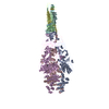
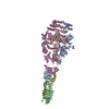
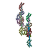
| Title | Structure of Francisella PdpA-VgrG Complex, Lidded | ||||||
 Components Components |
| ||||||
 Keywords Keywords | TRANSPORT PROTEIN / Type VI Secretion System / Complex | ||||||
| Function / homology | symbiont cell surface / host cell cytoplasm / Uncharacterized protein / Uncharacterized protein Function and homology information Function and homology information | ||||||
| Biological species |  Francisella tularensis subsp. novicida (bacteria) Francisella tularensis subsp. novicida (bacteria) | ||||||
| Method | ELECTRON MICROSCOPY / single particle reconstruction / cryo EM / Resolution: 4.35 Å | ||||||
 Authors Authors | Yang, X. / Clemens, D.L. / Lee, B.-Y. / Cui, Y.X. / Horwitz, M.A. / Zhou, Z.H. | ||||||
| Funding support |  United States, 1items United States, 1items
| ||||||
 Citation Citation |  Journal: Structure / Year: 2019 Journal: Structure / Year: 2019Title: Atomic Structure of the Francisella T6SS Central Spike Reveals a Unique α-Helical Lid and a Putative Cargo. Authors: Xue Yang / Daniel L Clemens / Bai-Yu Lee / Yanxiang Cui / Z Hong Zhou / Marcus A Horwitz /   Abstract: Francisella bacteria rely on a phylogenetically distinct type VI secretion system (T6SS) to escape host phagosomes and cause the fatal disease tularemia, but the structural and molecular mechanisms ...Francisella bacteria rely on a phylogenetically distinct type VI secretion system (T6SS) to escape host phagosomes and cause the fatal disease tularemia, but the structural and molecular mechanisms involved are unknown. Here we report the atomic structure of the Francisella T6SS central spike complex, obtained by cryo-electron microscopy. Our structural and functional studies demonstrate that, unlike the single-protein spike composition of other T6SS subtypes, Francisella T6SS's central spike is formed by two proteins, PdpA and VgrG, akin to T4-bacteriophage gp27 and gp5, respectively, and that PdpA has unique characteristics, including a putative cargo within its cavity and an N-terminal helical lid. Structure-guided mutagenesis demonstrates that the PdpA N-terminal lid and C-terminal spike are essential to Francisella T6SS function. PdpA is thus both an adaptor, connecting VgrG to the tube, and a likely carrier of secreted cargo. These findings are important to understanding Francisella pathogenicity and designing therapeutics to combat tularemia. | ||||||
| History |
|
- Structure visualization
| Movie |
 Movie viewer Movie viewer |
|---|---|
| Structure viewer | Molecule:  Molmil Molmil Jmol/JSmol Jmol/JSmol |
- Downloads & links
- Download
| PDBx/mmCIF format |  6u9f.cif.gz 6u9f.cif.gz | 436.2 KB | Display |  PDBx/mmCIF format PDBx/mmCIF format |
|---|---|---|---|---|
| PDB format |  pdb6u9f.ent.gz pdb6u9f.ent.gz | 349.6 KB | Display |  PDB format PDB format |
| PDBx/mmJSON format |  6u9f.json.gz 6u9f.json.gz | Tree view |  PDBx/mmJSON format PDBx/mmJSON format | |
| Others |  Other downloads Other downloads |
-Validation report
| Summary document |  6u9f_validation.pdf.gz 6u9f_validation.pdf.gz | 735.1 KB | Display |  wwPDB validaton report wwPDB validaton report |
|---|---|---|---|---|
| Full document |  6u9f_full_validation.pdf.gz 6u9f_full_validation.pdf.gz | 758.8 KB | Display | |
| Data in XML |  6u9f_validation.xml.gz 6u9f_validation.xml.gz | 65.1 KB | Display | |
| Data in CIF |  6u9f_validation.cif.gz 6u9f_validation.cif.gz | 101.8 KB | Display | |
| Arichive directory |  https://data.pdbj.org/pub/pdb/validation_reports/u9/6u9f https://data.pdbj.org/pub/pdb/validation_reports/u9/6u9f ftp://data.pdbj.org/pub/pdb/validation_reports/u9/6u9f ftp://data.pdbj.org/pub/pdb/validation_reports/u9/6u9f | HTTPS FTP |
-Related structure data
| Related structure data |  20696MC  6u9eC  6u9gC M: map data used to model this data C: citing same article ( |
|---|---|
| Similar structure data |
- Links
Links
- Assembly
| Deposited unit | 
|
|---|---|
| 1 |
|
- Components
| #1: Protein | Mass: 95469.961 Da / Num. of mol.: 3 Source method: isolated from a genetically manipulated source Source: (gene. exp.)  Francisella tularensis subsp. novicida (strain U112) (bacteria) Francisella tularensis subsp. novicida (strain U112) (bacteria)Strain: U112 / Gene: pdpA, FTN_1309 / Production host:  #2: Protein | Mass: 20539.779 Da / Num. of mol.: 3 Source method: isolated from a genetically manipulated source Source: (gene. exp.)  Francisella tularensis subsp. novicida (strain U112) (bacteria) Francisella tularensis subsp. novicida (strain U112) (bacteria)Strain: U112 / Gene: FTN_1312 / Production host:  |
|---|
-Experimental details
-Experiment
| Experiment | Method: ELECTRON MICROSCOPY |
|---|---|
| EM experiment | Aggregation state: PARTICLE / 3D reconstruction method: single particle reconstruction |
- Sample preparation
| Component | Name: PdpA-VgrG Complex / Type: COMPLEX / Entity ID: all / Source: RECOMBINANT |
|---|---|
| Source (natural) | Organism:  Francisella tularensis subsp. novicida U112 (bacteria) Francisella tularensis subsp. novicida U112 (bacteria) |
| Source (recombinant) | Organism:  |
| Buffer solution | pH: 7.5 |
| Specimen | Embedding applied: NO / Shadowing applied: NO / Staining applied: NO / Vitrification applied: YES |
| Specimen support | Grid material: COPPER / Grid mesh size: 300 divisions/in. / Grid type: Quantifoil R2/1 |
| Vitrification | Instrument: FEI VITROBOT MARK IV / Cryogen name: ETHANE / Humidity: 100 % / Chamber temperature: 298 K |
- Electron microscopy imaging
| Experimental equipment |  Model: Titan Krios / Image courtesy: FEI Company |
|---|---|
| Microscopy | Model: FEI TITAN KRIOS |
| Electron gun | Electron source:  FIELD EMISSION GUN / Accelerating voltage: 300 kV / Illumination mode: FLOOD BEAM FIELD EMISSION GUN / Accelerating voltage: 300 kV / Illumination mode: FLOOD BEAM |
| Electron lens | Mode: BRIGHT FIELD / Alignment procedure: COMA FREE |
| Specimen holder | Cryogen: NITROGEN / Specimen holder model: FEI TITAN KRIOS AUTOGRID HOLDER |
| Image recording | Electron dose: 47 e/Å2 / Detector mode: SUPER-RESOLUTION / Film or detector model: GATAN K2 SUMMIT (4k x 4k) |
- Processing
| EM software |
| ||||||||||||
|---|---|---|---|---|---|---|---|---|---|---|---|---|---|
| CTF correction | Type: PHASE FLIPPING ONLY | ||||||||||||
| Symmetry | Point symmetry: C3 (3 fold cyclic) | ||||||||||||
| 3D reconstruction | Resolution: 4.35 Å / Resolution method: FSC 0.143 CUT-OFF / Num. of particles: 3765 / Symmetry type: POINT | ||||||||||||
| Atomic model building | Space: REAL |
 Movie
Movie Controller


 PDBj
PDBj