 Movie
Movie Controller

[English] 日本語
 Yorodumi
- PDB-6u8y: Structure of the membrane-bound sulfane sulfur reductase (MBS), a... -
+ Open data

- Basic information
| Entry | Database: PDB / ID: 6u8y | ||||||||||||||||||||||||||||||||||||||||||||||||||||||
|---|---|---|---|---|---|---|---|---|---|---|---|---|---|---|---|---|---|---|---|---|---|---|---|---|---|---|---|---|---|---|---|---|---|---|---|---|---|---|---|---|---|---|---|---|---|---|---|---|---|---|---|---|---|---|---|
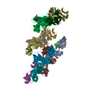
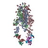
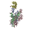
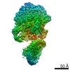
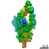
| Title | Structure of the membrane-bound sulfane sulfur reductase (MBS), an archaeal respiratory membrane complex | ||||||||||||||||||||||||||||||||||||||||||||||||||||||
 Components Components |
| ||||||||||||||||||||||||||||||||||||||||||||||||||||||
 Keywords Keywords | MEMBRANE PROTEIN / cryoEM / respiratory system | ||||||||||||||||||||||||||||||||||||||||||||||||||||||
| Function / homology |  Function and homology information Function and homology informationNADH dehydrogenase (quinone) / sodium:proton antiporter activity / monoatomic cation transmembrane transporter activity / electron transport coupled proton transport / oxidoreductase activity, acting on NAD(P)H / respiratory chain complex I / NADH dehydrogenase (ubiquinone) activity / quinone binding / aerobic respiration / NAD binding ...NADH dehydrogenase (quinone) / sodium:proton antiporter activity / monoatomic cation transmembrane transporter activity / electron transport coupled proton transport / oxidoreductase activity, acting on NAD(P)H / respiratory chain complex I / NADH dehydrogenase (ubiquinone) activity / quinone binding / aerobic respiration / NAD binding / 4 iron, 4 sulfur cluster binding / oxidoreductase activity / membrane / metal ion binding / plasma membrane Similarity search - Function | ||||||||||||||||||||||||||||||||||||||||||||||||||||||
| Biological species |   Pyrococcus furiosus COM1 (archaea) Pyrococcus furiosus COM1 (archaea) | ||||||||||||||||||||||||||||||||||||||||||||||||||||||
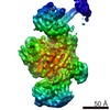
| Method | ELECTRON MICROSCOPY / single particle reconstruction / cryo EM / Resolution: 4 Å | ||||||||||||||||||||||||||||||||||||||||||||||||||||||
 Authors Authors | Yu, H.J. / Li, H.L. | ||||||||||||||||||||||||||||||||||||||||||||||||||||||
| Funding support |  United States, 2items United States, 2items
| ||||||||||||||||||||||||||||||||||||||||||||||||||||||
 Citation Citation | Journal: Nat Commun / Year: 2020 Title: Structure of the respiratory MBS complex reveals iron-sulfur cluster catalyzed sulfane sulfur reduction in ancient life. Authors: Hongjun Yu / Dominik K Haja / Gerrit J Schut / Chang-Hao Wu / Xing Meng / Gongpu Zhao / Huilin Li / Michael W W Adams /  Abstract: Modern day aerobic respiration in mitochondria involving complex I converts redox energy into chemical energy and likely evolved from a simple anaerobic system now represented by hydrogen gas- ...Modern day aerobic respiration in mitochondria involving complex I converts redox energy into chemical energy and likely evolved from a simple anaerobic system now represented by hydrogen gas-evolving hydrogenase (MBH) where protons are the terminal electron acceptor. Here we present the cryo-EM structure of an early ancestor in the evolution of complex I, the elemental sulfur (S)-reducing reductase MBS. Three highly conserved protein loops linking cytoplasmic and membrane domains enable scalable energy conversion in all three complexes. MBS contains two proton pumps compared to one in MBH and likely conserves twice the energy. The structure also reveals evolutionary adaptations of MBH that enabled S reduction by MBS catalyzed by a site-differentiated iron-sulfur cluster without participation of protons or amino acid residues. This is the simplest mechanism proposed for reduction of inorganic or organic disulfides. It is of fundamental significance in the iron and sulfur-rich volcanic environments of early earth and possibly the origin of life. MBS provides a new perspective on the evolution of modern-day respiratory complexes and of catalysis by biological iron-sulfur clusters. | ||||||||||||||||||||||||||||||||||||||||||||||||||||||
| History |
|
- Structure visualization
| Movie |
Movie viewer |
|---|---|
| Structure viewer | Molecule: MolmilJmol/JSmol |
- Downloads & links
-Download
| PDBx/mmCIF format | 6u8y.cif.gz | 1 MB | Display | PDBx/mmCIF format |
|---|---|---|---|---|
| PDB format | pdb6u8y.ent.gz | 861.1 KB | Display | PDB format |
| PDBx/mmJSON format | 6u8y.json.gz | Tree view | PDBx/mmJSON format | |
| Others |  Other downloads Other downloads |
-Validation report
| Arichive directory | https://data.pdbj.org/pub/pdb/validation_reports/u8/6u8yftp://data.pdbj.org/pub/pdb/validation_reports/u8/6u8y | HTTPS FTP |
|---|
-Related structure data
| Related structure data |  20692MC M: map data used to model this data C: citing same article ( |
|---|---|
| Similar structure data |

-Links
 PDBj
PDBj


- Assembly
| Deposited unit | 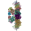
|
|---|---|
| 1 |
|
-Components
-Monovalent cation/H+ antiporter subunit ... , 5 types, 10 molecules AaBbCcEeGg
| #1: Protein | Mass: 19134.307 Da / Num. of mol.: 2 / Source method: isolated from a natural source / Source: (natural) Pyrococcus furiosus COM1 (archaea) / References: UniProt: I6UQN5#2: Protein | Mass: 9328.299 Da / Num. of mol.: 2 / Source method: isolated from a natural source / Source: (natural) Pyrococcus furiosus COM1 (archaea) / References: UniProt: I6UZW2#3: Protein | Mass: 13550.896 Da / Num. of mol.: 2 / Source method: isolated from a natural source / Source: (natural) Pyrococcus furiosus COM1 (archaea) / References: UniProt: I6TXQ5#5: Protein | Mass: 25691.111 Da / Num. of mol.: 2 / Source method: isolated from a natural source / Source: (natural) Pyrococcus furiosus COM1 (archaea) / References: UniProt: I6U860#6: Protein | Mass: 12073.443 Da / Num. of mol.: 2 / Source method: isolated from a natural source / Source: (natural) Pyrococcus furiosus COM1 (archaea) / References: UniProt: I6UQN0 |
|---|
-Protein , 2 types, 4 molecules DdMm
| #4: Protein | Mass: 10560.460 Da / Num. of mol.: 2 / Source method: isolated from a natural source / Source: (natural) Pyrococcus furiosus COM1 (archaea) / References: UniProt: I6V2A4#12: Protein | Mass: 33677.570 Da / Num. of mol.: 2 / Source method: isolated from a natural source / Source: (natural) Pyrococcus furiosus COM1 (archaea) / References: UniProt: I6V2A1 |
|---|
-NADH dehydrogenase subunit ... , 6 types, 12 molecules HhXxJjKkLlNn
| #7: Protein | Mass: 53019.332 Da / Num. of mol.: 2 / Source method: isolated from a natural source / Source: (natural) Pyrococcus furiosus COM1 (archaea) / References: UniProt: I6UZV7#8: Protein | Mass: 67561.906 Da / Num. of mol.: 2 / Source method: isolated from a natural source / Source: (natural) Pyrococcus furiosus COM1 (archaea) / References: UniProt: I6TXQ1#9: Protein | Mass: 22731.473 Da / Num. of mol.: 2 / Source method: isolated from a natural source / Source: (natural) Pyrococcus furiosus COM1 (archaea) / References: UniProt: I6UZV3, NADH dehydrogenase (quinone)#10: Protein | Mass: 20966.279 Da / Num. of mol.: 2 / Source method: isolated from a natural source / Source: (natural) Pyrococcus furiosus COM1 (archaea) / References: UniProt: I6TXP7, NADH dehydrogenase (quinone)#11: Protein | Mass: 44756.504 Da / Num. of mol.: 2 / Source method: isolated from a natural source / Source: (natural) Pyrococcus furiosus COM1 (archaea) / References: UniProt: I6V297, NADH dehydrogenase (quinone)#13: Protein | Mass: 24746.895 Da / Num. of mol.: 2 / Source method: isolated from a natural source / Source: (natural) Pyrococcus furiosus COM1 (archaea) / References: UniProt: I6U853, NADH dehydrogenase (quinone) |
|---|
-Non-polymers , 1 types, 6 molecules 
| #14: Chemical | ChemComp-SF4 /  Mass: 351.640 Da / Num. of mol.: 6 / Source method: obtained synthetically / Formula: Fe4S4 Mass: 351.640 Da / Num. of mol.: 6 / Source method: obtained synthetically / Formula: Fe4S4 |
|---|
-Details
| Has ligand of interest | N |
|---|---|
| Has protein modification | N |
-Experimental details
-Experiment
| Experiment | Method: ELECTRON MICROSCOPY |
|---|---|
| EM experiment | Aggregation state: PARTICLE / 3D reconstruction method: single particle reconstruction |
- Sample preparation
| Component | Name: respiratory membrane complex / Type: COMPLEX / Entity ID: #1-#13 / Source: NATURAL |
|---|---|
| Source (natural) | Organism: Pyrococcus furiosus (archaea) |
| Buffer solution | pH: 8.2 |
| Specimen | Embedding applied: NO / Shadowing applied: NO / Staining applied: NO / Vitrification applied: YES |
| Vitrification | Cryogen name: ETHANE |
- Electron microscopy imaging
| Experimental equipment |  Model: Titan Krios / Image courtesy: FEI Company |
|---|---|
| Microscopy | Model: FEI TITAN KRIOS |
| Electron gun | Electron source:  FIELD EMISSION GUN / Accelerating voltage: 300 kV / Illumination mode: FLOOD BEAM FIELD EMISSION GUN / Accelerating voltage: 300 kV / Illumination mode: FLOOD BEAM |
| Electron lens | Mode: BRIGHT FIELD |
| Image recording | Electron dose: 1.7 e/Å2 / Detector mode: COUNTING / Film or detector model: GATAN K2 SUMMIT (4k x 4k) |
- Processing
| Software | Name: PHENIX / Version: 1.16_3549: / Classification: refinement | ||||||||||||||||||||||||
|---|---|---|---|---|---|---|---|---|---|---|---|---|---|---|---|---|---|---|---|---|---|---|---|---|---|
| EM software |
| ||||||||||||||||||||||||
| CTF correction | Type: PHASE FLIPPING ONLY | ||||||||||||||||||||||||
| Symmetry | Point symmetry: C2 (2 fold cyclic) | ||||||||||||||||||||||||
| 3D reconstruction | Resolution: 4 Å / Resolution method: FSC 0.143 CUT-OFF / Num. of particles: 203673 / Symmetry type: POINT | ||||||||||||||||||||||||
| Refinement | Highest resolution: 4 Å | ||||||||||||||||||||||||
| Refine LS restraints |
|
