 Movie
Movie Controller

[English] 日本語
 Yorodumi
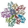
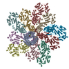
- PDB-6po2: In situ structure of BTV RNA-dependent RNA polymerase in BTV core -
+ Open data

- Basic information
| Entry | Database: PDB / ID: 6po2 | |||||||||||||||||||||||||||||||||
|---|---|---|---|---|---|---|---|---|---|---|---|---|---|---|---|---|---|---|---|---|---|---|---|---|---|---|---|---|---|---|---|---|---|---|
| Title | In situ structure of BTV RNA-dependent RNA polymerase in BTV core | |||||||||||||||||||||||||||||||||
 Components Components |
| |||||||||||||||||||||||||||||||||
 Keywords Keywords | VIRAL PROTEIN / TRANSFERASE / RNA dependent RNA polymerase | |||||||||||||||||||||||||||||||||
| Function / homology |  Function and homology information Function and homology informationviral genome replication / virion component / RNA-directed RNA polymerase / nucleotide binding / RNA-directed RNA polymerase activity / DNA-templated transcription / structural molecule activity / RNA binding Similarity search - Function | |||||||||||||||||||||||||||||||||
| Biological species |  Bluetongue virus 1 Bluetongue virus 1 | |||||||||||||||||||||||||||||||||
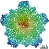
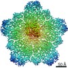
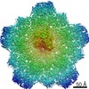
| Method | ELECTRON MICROSCOPY / single particle reconstruction / cryo EM / Resolution: 3.6 Å | |||||||||||||||||||||||||||||||||
 Authors Authors | He, Y. / Shivakoti, S. / Ding, K. / Cui, Y. / Roy, P. / Zhou, Z.H. | |||||||||||||||||||||||||||||||||
| Funding support |  United States, United States,  United Kingdom, 7items United Kingdom, 7items
| |||||||||||||||||||||||||||||||||
 Citation Citation | Journal: Proc Natl Acad Sci U S A / Year: 2019 Title: In situ structures of RNA-dependent RNA polymerase inside bluetongue virus before and after uncoating. Authors: Yao He / Sakar Shivakoti / Ke Ding / Yanxiang Cui / Polly Roy / Z Hong Zhou / Abstract: Bluetongue virus (BTV), a major threat to livestock, is a multilayered, nonturreted member of the , a family of segmented dsRNA viruses characterized by endogenous RNA transcription through an RNA- ...Bluetongue virus (BTV), a major threat to livestock, is a multilayered, nonturreted member of the , a family of segmented dsRNA viruses characterized by endogenous RNA transcription through an RNA-dependent RNA polymerase (RdRp). To date, the structure of BTV RdRp has been unknown, limiting our mechanistic understanding of BTV transcription and hindering rational drug design effort targeting this essential enzyme. Here, we report the in situ structures of BTV RdRp VP1 in both the triple-layered virion and double-layered core, as determined by cryo-electron microscopy (cryoEM) and subparticle reconstruction. BTV RdRp has 2 unique motifs not found in other viral RdRps: a fingernail, attached to the conserved fingers subdomain, and a bundle of 3 helices: 1 from the palm subdomain and 2 from the N-terminal domain. BTV RdRp VP1 is anchored to the inner surface of the capsid shell via 5 asymmetrically arranged N termini of the inner capsid shell protein VP3A around the 5-fold axis. The structural changes of RdRp VP1 and associated capsid shell proteins between BTV virions and cores suggest that the detachment of the outer capsid proteins VP2 and VP5 during viral entry induces both global movements of the inner capsid shell and local conformational changes of the N-terminal latch helix (residues 34 to 51) of 1 inner capsid shell protein VP3A, priming RdRp VP1 within the capsid for transcription. Understanding this mechanism in BTV also provides general insights into RdRp activation and regulation during viral entry of other multilayered, nonturreted dsRNA viruses. | |||||||||||||||||||||||||||||||||
| History |
|
- Structure visualization
| Movie |
Movie viewer |
|---|---|
| Structure viewer | Molecule: MolmilJmol/JSmol |
- Downloads & links
-Download
| PDBx/mmCIF format | 6po2.cif.gz | 1.7 MB | Display | PDBx/mmCIF format |
|---|---|---|---|---|
| PDB format | pdb6po2.ent.gz | 1.4 MB | Display | PDB format |
| PDBx/mmJSON format | 6po2.json.gz | Tree view | PDBx/mmJSON format | |
| Others |  Other downloads Other downloads |
-Validation report
| Arichive directory | https://data.pdbj.org/pub/pdb/validation_reports/po/6po2ftp://data.pdbj.org/pub/pdb/validation_reports/po/6po2 | HTTPS FTP |
|---|
-Related structure data
| Related structure data |  20407MC  6pnsC M: map data used to model this data C: citing same article ( |
|---|---|
| Similar structure data |


-Links
 PDBj
PDBj
- Assembly
| Deposited unit | 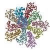
|
|---|---|
| 1 |
|
-Components
| #1: Protein | Mass: 149926.281 Da / Num. of mol.: 1 / Source method: isolated from a natural source / Source: (natural) Bluetongue virus 1 / References: UniProt: W0G557, RNA-directed RNA polymerase | ||
|---|---|---|---|
| #2: Protein | Mass: 103410.508 Da / Num. of mol.: 10 Source method: isolated from a genetically manipulated source Source: (gene. exp.) Bluetongue virus 1 / Gene: VP3 / Production host: Bluetongue virus 1 / References: UniProt: Q1AE73Has protein modification | N | |
-Experimental details
-Experiment
| Experiment | Method: ELECTRON MICROSCOPY |
|---|---|
| EM experiment | Aggregation state: PARTICLE / 3D reconstruction method: single particle reconstruction |
- Sample preparation
| Component | Name: Bluetongue virus 1 / Type: VIRUS / Entity ID: all / Source: NATURAL |
|---|---|
| Source (natural) | Organism: Bluetongue virus 1 |
| Details of virus | Empty: NO / Enveloped: NO / Isolate: SPECIES / Type: VIRION |
| Buffer solution | pH: 8.8 |
| Specimen | Embedding applied: NO / Shadowing applied: NO / Staining applied: NO / Vitrification applied: YES |
| Specimen support | Details: unspecified |
| Vitrification | Cryogen name: ETHANE |
- Electron microscopy imaging
| Experimental equipment |  Model: Titan Krios / Image courtesy: FEI Company |
|---|---|
| Microscopy | Model: FEI TITAN KRIOS |
| Electron gun | Electron source:  FIELD EMISSION GUN / Accelerating voltage: 300 kV / Illumination mode: FLOOD BEAM FIELD EMISSION GUN / Accelerating voltage: 300 kV / Illumination mode: FLOOD BEAM |
| Electron lens | Mode: BRIGHT FIELD / Cs: 2.7 mm |
| Image recording | Electron dose: 32 e/Å2 / Detector mode: SUPER-RESOLUTION / Film or detector model: GATAN K2 SUMMIT (4k x 4k) |
- Processing
| Software | Name: PHENIX / Version: 1.14_3260: / Classification: refinement |
|---|---|
| EM software | Name: PHENIX / Category: model refinement |
| CTF correction | Type: PHASE FLIPPING AND AMPLITUDE CORRECTION |
| Symmetry | Point symmetry: C1 (asymmetric) |
| 3D reconstruction | Resolution: 3.6 Å / Resolution method: FSC 0.143 CUT-OFF / Num. of particles: 150346 / Symmetry type: POINT |
