 Movie
Movie Controller


+ Open data

- Basic information
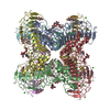
| Entry | Database: PDB / ID: 6lyg | ||||||||||||||||||
|---|---|---|---|---|---|---|---|---|---|---|---|---|---|---|---|---|---|---|---|
| Title | Cryo-EM structure of the calcium homeostasis modulator 1 channel | ||||||||||||||||||
 Components Components | Calcium homeostasis modulator 1 | ||||||||||||||||||
 Keywords Keywords | MEMBRANE PROTEIN / Octamer | ||||||||||||||||||
| Function / homology | Calcium homeostasis modulator family / Calcium homeostasis modulator / ATP export / voltage-gated calcium channel activity / plasma membrane / Calcium homeostasis modulator 1 Function and homology information Function and homology information | ||||||||||||||||||
| Biological species |  | ||||||||||||||||||
| Method | ELECTRON MICROSCOPY / single particle reconstruction / cryo EM / Resolution: 3.1 Å | ||||||||||||||||||
 Authors Authors | Ren, Y. / Yang, X. / Shen, Y. | ||||||||||||||||||
| Funding support |  China, 5items China, 5items
| ||||||||||||||||||
 Citation Citation | Journal: Sci Adv / Year: 2020 Title: Cryo-EM structure of the calcium homeostasis modulator 1 channel. Authors: Yue Ren / Tianlei Wen / Zhiqin Xi / Shunjin Li / Jing Lu / Xing Zhang / Xue Yang / Yuequan Shen / Abstract: Calcium homeostasis modulator 1 (CALHM1) is a voltage-gated ATP release channel that plays an important role in neural gustatory signaling and the pathogenesis of Alzheimer's disease. Here, we ...Calcium homeostasis modulator 1 (CALHM1) is a voltage-gated ATP release channel that plays an important role in neural gustatory signaling and the pathogenesis of Alzheimer's disease. Here, we present a cryo-electron microscopy structure of full-length Ca-free CALHM1 from Danio rerio at an overall resolution of 3.1 Å. Our structure reveals an octameric architecture with a wide pore diameter of ~20 Å, presumably representing the active conformation. The overall structure is substantially different from that of the isoform CALHM2, which forms both undecameric hemichannels and gap junctions. The N-terminal small helix folds back to the pore and forms an antiparallel interaction with transmembrane helix 1. Structural analysis revealed that the extracellular loop 1 region within the dimer interface may contribute to oligomeric assembly. A positive potential belt inside the pore was identified that may modulate ion permeation. Our structure offers insights into the assembly and gating mechanism of the CALHM1 channel. | ||||||||||||||||||
| History |
|
- Structure visualization
| Movie |
Movie viewer |
|---|---|
| Structure viewer | Molecule: MolmilJmol/JSmol |
- Downloads & links
-Download
| PDBx/mmCIF format | 6lyg.cif.gz | 336.5 KB | Display | PDBx/mmCIF format |
|---|---|---|---|---|
| PDB format | pdb6lyg.ent.gz | 269.8 KB | Display | PDB format |
| PDBx/mmJSON format | 6lyg.json.gz | Tree view | PDBx/mmJSON format | |
| Others |  Other downloads Other downloads |
-Validation report
| Arichive directory | https://data.pdbj.org/pub/pdb/validation_reports/ly/6lygftp://data.pdbj.org/pub/pdb/validation_reports/ly/6lyg | HTTPS FTP |
|---|
-Related structure data
| Related structure data |  30016MC M: map data used to model this data C: citing same article ( |
|---|---|
| Similar structure data |


-Links
 PDBj
PDBj- Assembly
| Deposited unit | 
|
|---|---|
| 1 |
|
-Components
| #1: Protein | Mass: 41213.645 Da / Num. of mol.: 8 Source method: isolated from a genetically manipulated source Source: (gene. exp.)  Homo sapiens (human) / References: UniProt: E7F2J4 Homo sapiens (human) / References: UniProt: E7F2J4#2: Sugar | ChemComp-NAG /   Type: D-saccharide, beta linking / Mass: 221.208 Da / Num. of mol.: 8 Type: D-saccharide, beta linking / Mass: 221.208 Da / Num. of mol.: 8Source method: isolated from a genetically manipulated source Formula: C8H15NO6 Has ligand of interest | N | Has protein modification | Y | |
|---|
-Experimental details
-Experiment
| Experiment | Method: ELECTRON MICROSCOPY |
|---|---|
| EM experiment | Aggregation state: PARTICLE / 3D reconstruction method: single particle reconstruction |
- Sample preparation
| Component | Name: CALHM1 channel / Type: CELL / Entity ID: #1 / Source: RECOMBINANT |
|---|---|
| Source (natural) | Organism: |
| Source (recombinant) | Organism: Homo sapiens (human) |
| Buffer solution | pH: 7.5 |
| Specimen | Conc.: 8 mg/ml / Embedding applied: NO / Shadowing applied: NO / Staining applied: NO / Vitrification applied: YES |
| Vitrification | Instrument: FEI VITROBOT MARK IV / Cryogen name: ETHANE |
- Electron microscopy imaging
| Experimental equipment |  Model: Titan Krios / Image courtesy: FEI Company |
|---|---|
| Microscopy | Model: FEI TITAN KRIOS |
| Electron gun | Electron source:  FIELD EMISSION GUN / Accelerating voltage: 300 kV / Illumination mode: FLOOD BEAM FIELD EMISSION GUN / Accelerating voltage: 300 kV / Illumination mode: FLOOD BEAM |
| Electron lens | Mode: BRIGHT FIELD / Alignment procedure: COMA FREE |
| Specimen holder | Cryogen: NITROGEN / Specimen holder model: FEI TITAN KRIOS AUTOGRID HOLDER |
| Image recording | Electron dose: 50 e/Å2 / Detector mode: COUNTING / Film or detector model: GATAN K2 SUMMIT (4k x 4k) |
- Processing
| CTF correction | Type: NONE | ||||||||||||||||||||||||
|---|---|---|---|---|---|---|---|---|---|---|---|---|---|---|---|---|---|---|---|---|---|---|---|---|---|
| 3D reconstruction | Resolution: 3.1 Å / Resolution method: FSC 0.143 CUT-OFF / Num. of particles: 59833 / Symmetry type: POINT | ||||||||||||||||||||||||
| Refinement | Stereochemistry target values: GeoStd + Monomer Library + CDL v1.2 | ||||||||||||||||||||||||
| Displacement parameters | Biso mean: 125.59 Å2 | ||||||||||||||||||||||||
| Refine LS restraints |
|
