 Movie
Movie Controller

[English] 日本語
 Yorodumi
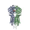
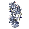
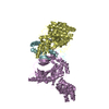
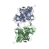
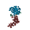
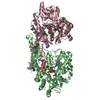
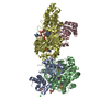
- PDB-6hbu: Cryo-EM structure of the ABCG2 E211Q mutant bound to ATP and Magnesium -
+ Open data

- Basic information
| Entry | Database: PDB / ID: 6hbu | |||||||||
|---|---|---|---|---|---|---|---|---|---|---|
| Title | Cryo-EM structure of the ABCG2 E211Q mutant bound to ATP and Magnesium | |||||||||
 Components Components | ATP-binding cassette sub-family G member 2 | |||||||||
 Keywords Keywords | MEMBRANE PROTEIN / Multidrug transporter / cancer | |||||||||
| Function / homology |  Function and homology information Function and homology informationbiotin transmembrane transporter activity / biotin transport / riboflavin transport / riboflavin transmembrane transporter activity / sphingolipid transporter activity / external side of apical plasma membrane / renal urate salt excretion / urate transmembrane transporter activity / urate metabolic process / Abacavir transmembrane transport ...biotin transmembrane transporter activity / biotin transport / riboflavin transport / riboflavin transmembrane transporter activity / sphingolipid transporter activity / external side of apical plasma membrane / renal urate salt excretion / urate transmembrane transporter activity / urate metabolic process / Abacavir transmembrane transport / organic anion transport / sphingolipid biosynthetic process / Sphingolipid de novo biosynthesis / organic anion transmembrane transporter activity / xenobiotic transport across blood-brain barrier / transepithelial transport / export across plasma membrane / ABC-type xenobiotic transporter / Paracetamol ADME / ABC-type xenobiotic transporter activity / Ciprofloxacin ADME / NFE2L2 regulating MDR associated enzymes / Differentiation of keratinocytes in interfollicular epidermis in mammalian skin / cellular detoxification / Heme biosynthesis / Heme degradation / xenobiotic transmembrane transporter activity / efflux transmembrane transporter activity / transport across blood-brain barrier / ATPase-coupled transmembrane transporter activity / mitochondrial membrane / brush border membrane / Iron uptake and transport / transmembrane transport / apical plasma membrane / membrane raft / protein homodimerization activity / ATP hydrolysis activity / nucleoplasm / ATP binding / identical protein binding / plasma membrane Similarity search - Function | |||||||||
| Biological species |  Homo sapiens (human) Homo sapiens (human) | |||||||||
| Method | ELECTRON MICROSCOPY / single particle reconstruction / cryo EM / Resolution: 3.09 Å | |||||||||
 Authors Authors | Manolaridis, I. / Jackson, S.M. / Taylor, N.M.I. / Kowal, J. / Stahlberg, H. / Locher, K.P. | |||||||||
| Funding support |  Switzerland, 2items Switzerland, 2items
| |||||||||
 Citation Citation | Journal: Nature / Year: 2018 Title: Cryo-EM structures of a human ABCG2 mutant trapped in ATP-bound and substrate-bound states. Authors: Ioannis Manolaridis / Scott M Jackson / Nicholas M I Taylor / Julia Kowal / Henning Stahlberg / Kaspar P Locher /  Abstract: ABCG2 is a transporter protein of the ATP-binding-cassette (ABC) family that is expressed in the plasma membrane in cells of various tissues and tissue barriers, including the blood-brain, blood- ...ABCG2 is a transporter protein of the ATP-binding-cassette (ABC) family that is expressed in the plasma membrane in cells of various tissues and tissue barriers, including the blood-brain, blood-testis and maternal-fetal barriers. Powered by ATP, it translocates endogenous substrates, affects the pharmacokinetics of many drugs and protects against a wide array of xenobiotics, including anti-cancer drugs. Previous studies have revealed the architecture of ABCG2 and the structural basis of its inhibition by small molecules and antibodies. However, the mechanisms of substrate recognition and ATP-driven transport are unknown. Here we present high-resolution cryo-electron microscopy (cryo-EM) structures of human ABCG2 in a substrate-bound pre-translocation state and an ATP-bound post-translocation state. For both structures, we used a mutant containing a glutamine replacing the catalytic glutamate (ABCG2), which resulted in reduced ATPase and transport rates and facilitated conformational trapping for structural studies. In the substrate-bound state, a single molecule of estrone-3-sulfate (ES) is bound in a central, hydrophobic and cytoplasm-facing cavity about halfway across the membrane. Only one molecule of ES can bind in the observed binding mode. In the ATP-bound state, the substrate-binding cavity has collapsed while an external cavity has opened to the extracellular side of the membrane. The ATP-induced conformational changes include rigid-body shifts of the transmembrane domains, pivoting of the nucleotide-binding domains (NBDs), and a change in the relative orientation of the NBD subdomains. Mutagenesis and in vitro characterization of transport and ATPase activities demonstrate the roles of specific residues in substrate recognition, including a leucine residue that forms a 'plug' between the two cavities. Our results show how ABCG2 harnesses the energy of ATP binding to extrude ES and other substrates, and suggest that the size and binding affinity of compounds are important for distinguishing substrates from inhibitors. | |||||||||
| History |
|
- Structure visualization
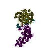
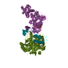
| Movie |
Movie viewer |
|---|---|
| Structure viewer | Molecule: MolmilJmol/JSmol |
- Downloads & links
-Download
| PDBx/mmCIF format | 6hbu.cif.gz | 206.9 KB | Display | PDBx/mmCIF format |
|---|---|---|---|---|
| PDB format | pdb6hbu.ent.gz | 170.8 KB | Display | PDB format |
| PDBx/mmJSON format | 6hbu.json.gz | Tree view | PDBx/mmJSON format | |
| Others |  Other downloads Other downloads |
-Validation report
| Summary document | 6hbu_validation.pdf.gz | 929.6 KB | Display | wwPDB validaton report |
|---|---|---|---|---|
| Full document | 6hbu_full_validation.pdf.gz | 938.8 KB | Display | |
| Data in XML | 6hbu_validation.xml.gz | 40.8 KB | Display | |
| Data in CIF | 6hbu_validation.cif.gz | 61.8 KB | Display | |
| Arichive directory | https://data.pdbj.org/pub/pdb/validation_reports/hb/6hbuftp://data.pdbj.org/pub/pdb/validation_reports/hb/6hbu | HTTPS FTP |
-Related structure data
| Related structure data | 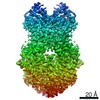 0190MC 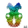 0196C 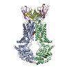 6hcoC  6hzmC M: map data used to model this data C: citing same article ( |
|---|---|
| Similar structure data |

-Links
 PDBj
PDBj


- Assembly
| Deposited unit | 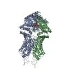
|
|---|---|
| 1 |
|
-Components
| #1: Protein | Mass: 72384.867 Da / Num. of mol.: 2 / Mutation: E211Q Source method: isolated from a genetically manipulated source Source: (gene. exp.) Homo sapiens (human) / Gene: ABCG2, ABCP, BCRP, BCRP1, MXR / Cell line (production host): HEK293 EBNA / Production host: Homo sapiens (human) / References: UniProt: Q9UNQ0#2: Chemical |   Mass: 507.181 Da / Num. of mol.: 2 / Source method: obtained synthetically / Formula: C10H16N5O13P3 / Comment: ATP, energy-carrying molecule*YM Mass: 507.181 Da / Num. of mol.: 2 / Source method: obtained synthetically / Formula: C10H16N5O13P3 / Comment: ATP, energy-carrying molecule*YM#3: Chemical |   Mass: 24.305 Da / Num. of mol.: 2 / Source method: obtained synthetically / Formula: Mg Mass: 24.305 Da / Num. of mol.: 2 / Source method: obtained synthetically / Formula: Mg |
|---|
-Experimental details
-Experiment
| Experiment | Method: ELECTRON MICROSCOPY |
|---|---|
| EM experiment | Aggregation state: PARTICLE / 3D reconstruction method: single particle reconstruction |
- Sample preparation
| Component |
| ||||||||||||||||||
|---|---|---|---|---|---|---|---|---|---|---|---|---|---|---|---|---|---|---|---|
| Source (natural) | Organism: HOMO SAPIENS (human) | ||||||||||||||||||
| Source (recombinant) | Organism: HOMO SAPIENS (human) | ||||||||||||||||||
| Buffer solution | pH: 7.5 | ||||||||||||||||||
| Specimen | Conc.: 0.4 mg/ml / Embedding applied: NO / Shadowing applied: NO / Staining applied: NO / Vitrification applied: YES | ||||||||||||||||||
| Specimen support | Grid material: COPPER / Grid mesh size: 300 divisions/in. / Grid type: Quantifoil R1.2/1.3 | ||||||||||||||||||
| Vitrification | Instrument: FEI VITROBOT MARK IV / Cryogen name: ETHANE-PROPANE / Humidity: 100 % / Chamber temperature: 277 K |
- Electron microscopy imaging
| Experimental equipment |  Model: Titan Krios / Image courtesy: FEI Company |
|---|---|
| Microscopy | Model: FEI TITAN KRIOS |
| Electron gun | Electron source:  FIELD EMISSION GUN / Accelerating voltage: 300 kV / Illumination mode: FLOOD BEAM FIELD EMISSION GUN / Accelerating voltage: 300 kV / Illumination mode: FLOOD BEAM |
| Electron lens | Mode: BRIGHT FIELD / Nominal magnification: 165000 X |
| Image recording | Average exposure time: 0.2 sec. / Electron dose: 2 e/Å2 / Film or detector model: GATAN K2 SUMMIT (4k x 4k) / Num. of real images: 4905 |
- Processing
| Software | Name: PHENIX / Version: 1.13_2998: / Classification: refinement | ||||||||||||||||||||||||
|---|---|---|---|---|---|---|---|---|---|---|---|---|---|---|---|---|---|---|---|---|---|---|---|---|---|
| EM software |
| ||||||||||||||||||||||||
| CTF correction | Type: PHASE FLIPPING AND AMPLITUDE CORRECTION | ||||||||||||||||||||||||
| Symmetry | Point symmetry: C1 (asymmetric) | ||||||||||||||||||||||||
| 3D reconstruction | Resolution: 3.09 Å / Resolution method: FSC 0.143 CUT-OFF / Num. of particles: 288447 / Symmetry type: POINT | ||||||||||||||||||||||||
| Atomic model building | B value: 136 | ||||||||||||||||||||||||
| Refine LS restraints |
|
