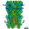
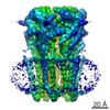
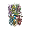
ジャーナル: Cell / 年: 2016 タイトル: Structure of a Chaperone-Usher Pilus Reveals the Molecular Basis of Rod Uncoiling. 著者: Manuela K Hospenthal / Adam Redzej / Karen Dodson / Marta Ukleja / Brandon Frenz / Catarina Rodrigues / Scott J Hultgren / Frank DiMaio / Edward H Egelman / Gabriel Waksman / 要旨: Types 1 and P pili are prototypical bacterial cell-surface appendages playing essential roles in mediating adhesion of bacteria to the urinary tract. These pili, assembled by the chaperone-usher ...Types 1 and P pili are prototypical bacterial cell-surface appendages playing essential roles in mediating adhesion of bacteria to the urinary tract. These pili, assembled by the chaperone-usher pathway, are polymers of pilus subunits assembling into two parts: a thin, short tip fibrillum at the top, mounted on a long pilus rod. The rod adopts a helical quaternary structure and is thought to play essential roles: its formation may drive pilus extrusion by preventing backsliding of the nascent growing pilus within the secretion pore; the rod also has striking spring-like properties, being able to uncoil and recoil depending on the intensity of shear forces generated by urine flow. Here, we present an atomic model of the P pilus generated from a 3.8 Å resolution cryo-electron microscopy reconstruction. This structure provides the molecular basis for the rod's remarkable mechanical properties and illuminates its role in pilus secretion.
A: PAP FIMBRIAL MAJOR PILIN PROTEIN B: PAP FIMBRIAL MAJOR PILIN PROTEIN C: PAP FIMBRIAL MAJOR PILIN PROTEIN D: PAP FIMBRIAL MAJOR PILIN PROTEIN E: PAP FIMBRIAL MAJOR PILIN PROTEIN F: PAP FIMBRIAL MAJOR PILIN PROTEIN G: PAP FIMBRIAL MAJOR PILIN PROTEIN H: PAP FIMBRIAL MAJOR PILIN PROTEIN I: PAP FIMBRIAL MAJOR PILIN PROTEIN J: PAP FIMBRIAL MAJOR PILIN PROTEIN K: PAP FIMBRIAL MAJOR PILIN PROTEIN L: PAP FIMBRIAL MAJOR PILIN PROTEIN M: PAP FIMBRIAL MAJOR PILIN PROTEIN
モード: BRIGHT FIELD / 最大 デフォーカス(公称値): 3000 nm / 最小 デフォーカス(公称値): 1000 nm / Cs: 2.7 mm
撮影
電子線照射量: 17 e/Å2 フィルム・検出器のモデル: FEI FALCON II (4k x 4k)
画像スキャン
デジタル画像の数: 90
-
解析
EMソフトウェア
名称: SPIDER / カテゴリ: 3次元再構成
CTF補正
詳細: EACH IMAGE
3次元再構成
手法: IHRSR / 解像度: 3.8 Å / 粒子像の数: 56341 / ピクセルサイズ(公称値): 1.1 Å / ピクセルサイズ(実測値): 1.1 Å 詳細: SUBMISSION BASED ON EXPERIMENTAL DATA FROM EMDB EMD-3222. (DEPOSITION ID: 13990). 対称性のタイプ: HELICAL
原子モデル構築
プロトコル: OTHER / 空間: REAL / 詳細: REFINEMENT PROTOCOL--EM
 ムービー
ムービー コントローラー


 データを開く
 基本情報
基本情報 要素
要素 キーワード
キーワード 機能・相同性情報
 データ登録者
 引用
引用

 構造の表示
構造の表示 ダウンロードとリンク
 その他のダウンロード


 PDBj
PDBj 集合体
集合体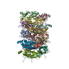
 試料調製
試料調製 電子顕微鏡撮影
 FIELD EMISSION GUN / 加速電圧: 300 kV / 照射モード: FLOOD BEAM
 解析
解析