+ Open data
- Basic information
| Entry | Database: PDB / ID: 5an8 | ||||||
|---|---|---|---|---|---|---|---|
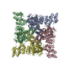
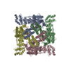
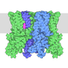
| Title | Cryo-electron microscopy structure of rabbit TRPV2 ion channel | ||||||
 Components Components | TRPV2 | ||||||
 Keywords Keywords | TRANSPORT PROTEIN / TRP CHANNEL | ||||||
| Function / homology |  Function and homology information Function and homology informationgrowth cone membrane / response to temperature stimulus / positive regulation of calcium ion import / calcium ion import across plasma membrane / positive regulation of axon extension / axonal growth cone / calcium channel activity / positive regulation of cold-induced thermogenesis / cell body / cell surface / identical protein binding Similarity search - Function | ||||||
| Biological species |  | ||||||
| Method | ELECTRON MICROSCOPY / single particle reconstruction / cryo EM / Resolution: 3.8 Å | ||||||
 Authors Authors | Zubcevic, L. / Herzik, M.A.J. / Chung, B.C. / Lander, G.C. / Lee, S.Y. | ||||||
 Citation Citation |  Journal: Nat Struct Mol Biol / Year: 2016 Journal: Nat Struct Mol Biol / Year: 2016Title: Cryo-electron microscopy structure of the TRPV2 ion channel. Authors: Lejla Zubcevic / Mark A Herzik / Ben C Chung / Zhirui Liu / Gabriel C Lander / Seok-Yong Lee /  Abstract: Transient receptor potential vanilloid (TRPV) cation channels are polymodal sensors involved in a variety of physiological processes. TRPV2, a member of the TRPV family, is regulated by temperature, ...Transient receptor potential vanilloid (TRPV) cation channels are polymodal sensors involved in a variety of physiological processes. TRPV2, a member of the TRPV family, is regulated by temperature, by ligands, such as probenecid and cannabinoids, and by lipids. TRPV2 has been implicated in many biological functions, including somatosensation, osmosensation and innate immunity. Here we present the atomic model of rabbit TRPV2 in its putative desensitized state, as determined by cryo-EM at a nominal resolution of ∼4 Å. In the TRPV2 structure, the transmembrane segment 6 (S6), which is involved in gate opening, adopts a conformation different from the one observed in TRPV1. Structural comparisons of TRPV1 and TRPV2 indicate that a rotation of the ankyrin-repeat domain is coupled to pore opening via the TRP domain, and this pore opening can be modulated by rearrangements in the secondary structure of S6. | ||||||
| History |
|
- Structure visualization
| Movie |
 Movie viewer Movie viewer |
|---|---|
| Structure viewer | Molecule:  Molmil Molmil Jmol/JSmol Jmol/JSmol |
- Downloads & links
- Download
| PDBx/mmCIF format |  5an8.cif.gz 5an8.cif.gz | 419.5 KB | Display |  PDBx/mmCIF format PDBx/mmCIF format |
|---|---|---|---|---|
| PDB format |  pdb5an8.ent.gz pdb5an8.ent.gz | 327.3 KB | Display |  PDB format PDB format |
| PDBx/mmJSON format |  5an8.json.gz 5an8.json.gz | Tree view |  PDBx/mmJSON format PDBx/mmJSON format | |
| Others |  Other downloads Other downloads |
-Validation report
| Arichive directory |  https://data.pdbj.org/pub/pdb/validation_reports/an/5an8 https://data.pdbj.org/pub/pdb/validation_reports/an/5an8 ftp://data.pdbj.org/pub/pdb/validation_reports/an/5an8 ftp://data.pdbj.org/pub/pdb/validation_reports/an/5an8 | HTTPS FTP |
|---|
-Related structure data
| Related structure data |  6455MC M: map data used to model this data C: citing same article ( |
|---|---|
| Similar structure data |
- Links
Links
- Assembly
| Deposited unit | 
|
|---|---|
| 1 |
|
- Components
| #1: Protein | Mass: 69906.391 Da / Num. of mol.: 4 Source method: isolated from a genetically manipulated source Details: RABBIT TRPV2 / Source: (gene. exp.)   Has protein modification | Y | |
|---|
-Experimental details
-Experiment
| Experiment | Method: ELECTRON MICROSCOPY |
|---|---|
| EM experiment | Aggregation state: PARTICLE / 3D reconstruction method: single particle reconstruction |
- Sample preparation
| Component | Name: RABBIT TRPV2 / Type: COMPLEX |
|---|---|
| Buffer solution | Name: 20MM PBS / pH: 7.6 / Details: 20MM PBS |
| Specimen | Conc.: 1 mg/ml / Embedding applied: NO / Shadowing applied: NO / Staining applied: NO / Vitrification applied: YES |
| Specimen support | Details: HOLEY CARBON |
| Vitrification | Instrument: GATAN CRYOPLUNGE 3 / Cryogen name: ETHANE / Details: PLUNGE FROZEN IN LIQUID ETHANE |
- Electron microscopy imaging
| Experimental equipment |  Model: Titan Krios / Image courtesy: FEI Company |
|---|---|
| Microscopy | Model: FEI TITAN KRIOS / Date: May 12, 2015 Details: DATA ACQUIRED USING LEGINON, COLLECTED IN K2 SUPER RESOLUTION MODE |
| Electron gun | Electron source:  FIELD EMISSION GUN / Accelerating voltage: 300 kV / Illumination mode: FLOOD BEAM FIELD EMISSION GUN / Accelerating voltage: 300 kV / Illumination mode: FLOOD BEAM |
| Electron lens | Mode: BRIGHT FIELD / Nominal magnification: 22500 X / Calibrated magnification: 38168 X / Nominal defocus max: 3000 nm / Nominal defocus min: 1200 nm / Cs: 2.7 mm |
| Specimen holder | Temperature: 78 K |
| Image recording | Electron dose: 57 e/Å2 / Film or detector model: GATAN K2 SUMMIT (4k x 4k) |
| Image scans | Num. digital images: 747 |
- Processing
| Symmetry | Point symmetry: C4 (4 fold cyclic) | ||||||||||||
|---|---|---|---|---|---|---|---|---|---|---|---|---|---|
| 3D reconstruction | Resolution: 3.8 Å / Resolution method: FSC 0.143 CUT-OFF / Num. of particles: 43879 / Refinement type: HALF-MAPS REFINED INDEPENDENTLY / Symmetry type: POINT | ||||||||||||
| Refinement | Highest resolution: 3.8 Å | ||||||||||||
| Refinement step | Cycle: LAST / Highest resolution: 3.8 Å
|
 Movie
Movie Controller


 PDBj
PDBj
