[English] 日本語
Yorodumi
- PDB-4v1a: Structure of the large subunit of the mammalian mitoribosome, par... -

+
Open data


ID or keywords:

Loading...

-
Basic information

Entry
Database: PDB / ID: 4v1a
TitleStructure of the large subunit of the mammalian mitoribosome, part 2 of 2
Components
  • (MITORIBOSOMAL PROTEIN ...) x 22
  • UNASSIGNED SECONDARY STRUCTURE ELEMENTS
KeywordsRIBOSOME / TRANSLATION / LARGE RIBOSOMAL SUBUNIT / MITORIBOSOME / MAMMALIAN MITOCHONDRIAL RIBOSOME / CRYO-EM
Function / homology
Function and homology information


Mitochondrial translation elongation / Mitochondrial translation termination / translation release factor activity / rRNA import into mitochondrion / mitochondrial translational elongation / ribonuclease III activity / microprocessor complex / mitochondrial large ribosomal subunit / peptidyl-tRNA hydrolase / mitochondrial ribosome ...Mitochondrial translation elongation / Mitochondrial translation termination / translation release factor activity / rRNA import into mitochondrion / mitochondrial translational elongation / ribonuclease III activity / microprocessor complex / mitochondrial large ribosomal subunit / peptidyl-tRNA hydrolase / mitochondrial ribosome / mitochondrial translation / RNA processing / double-stranded RNA binding / cell junction / 5S rRNA binding / nuclear body / ribosome / structural constituent of ribosome / ribonucleoprotein complex / translation / nucleotide binding / mitochondrion / nucleoplasm / nucleus / plasma membrane / cytosol
Similarity search - Function
Single alpha-helices involved in coiled-coils or other helix-helix interfaces - #3440 / Growth arrest and DNA-damage-inducible proteins-interacting protein 1 / Coiled-coil domain-containing protein 142 / Coiled-coil protein 142 / SUI1-like domain / Translation Initiation Factor Eif1 / Mitochondrial ribosomal protein L48 / Mitochondrial ribosomal protein L46 NUDIX / 39S ribosomal protein L52, mitochondrial / Mitoribosomal protein mL52 ...Single alpha-helices involved in coiled-coils or other helix-helix interfaces - #3440 / Growth arrest and DNA-damage-inducible proteins-interacting protein 1 / Coiled-coil domain-containing protein 142 / Coiled-coil protein 142 / SUI1-like domain / Translation Initiation Factor Eif1 / Mitochondrial ribosomal protein L48 / Mitochondrial ribosomal protein L46 NUDIX / 39S ribosomal protein L52, mitochondrial / Mitoribosomal protein mL52 / : / Methane Monooxygenase Hydroxylase; Chain G, domain 1 / MRPL44, double-stranded RNA binding domain / MRPL44 dsRNA-binding domain / Large ribosomal subunit protein mL44, endonuclease domain / : / Ribosomal protein L46, N-terminal / 39S mitochondrial ribosomal protein L46 / Ribosomal protein L50, mitochondria / Ribosomal subunit 39S / 39S ribosomal protein L46, mitochondrial / 39S ribosomal protein L43/54S ribosomal protein L51 / Phosphatidylethanolamine-binding protein / Phosphatidylethanolamine-binding protein / Phosphatidylethanolamine-binding protein, eukaryotic / PEBP-like superfamily / Threonyl/alanyl tRNA synthetase, class II-like, putative editing domain superfamily / TGS-like / TGS domain profile. / TGS / Peptide chain release factor class I / RF-1 domain / Ribonuclease III, endonuclease domain superfamily / Double stranded RNA-binding domain (dsRBD) profile. / Nucleoside Triphosphate Pyrophosphohydrolase / Nucleoside Triphosphate Pyrophosphohydrolase / Double-stranded RNA-binding domain / Single alpha-helices involved in coiled-coils or other helix-helix interfaces / Beta-grasp domain superfamily / Ribosomal protein/NADH dehydrogenase domain / Mitochondrial ribosomal protein L51 / S25 / CI-B8 domain / Mitochondrial ribosomal protein L51 / S25 / CI-B8 domain / NUDIX hydrolase-like domain superfamily / Helix non-globular / Ribosomal protein L18 / Ribosomal L18 of archaea, bacteria, mitoch. and chloroplast / Special / Glutaredoxin / Ribosomal protein S18 / Ribosomal protein S18 / Ribosomal protein S18 superfamily / Glutaredoxin / Ribosomal protein S10p/S20e / Ribosomal protein S10 domain / Ribosomal protein S10 domain superfamily / Ribosomal protein S10p/S20e / Ribosomal protein S11 superfamily / Thioredoxin-like superfamily / Alpha-Beta Complex / 2-Layer Sandwich / 3-Layer(aba) Sandwich / Alpha Beta
Similarity search - Domain/homology
39S ribosomal protein L18, mitochondrial isoform 1 / 39S ribosomal protein L48, mitochondrial / : / Mitochondrial ribosomal protein S18A / Large ribosomal subunit protein mL38 / Large ribosomal subunit protein mL43 / Large ribosomal subunit protein mL52 / : / Mitochondrial ribosomal protein L39 / Coiled-coil domain containing 142 ...39S ribosomal protein L18, mitochondrial isoform 1 / 39S ribosomal protein L48, mitochondrial / : / Mitochondrial ribosomal protein S18A / Large ribosomal subunit protein mL38 / Large ribosomal subunit protein mL43 / Large ribosomal subunit protein mL52 / : / Mitochondrial ribosomal protein L39 / Coiled-coil domain containing 142 / Large ribosomal subunit protein mL44 / Large ribosomal subunit protein mL46 / Large ribosomal subunit protein mL50 / Large ribosomal subunit protein mL62
Similarity search - Component
Biological speciesSUS SCROFA (pig)
MethodELECTRON MICROSCOPY / single particle reconstruction / cryo EM / Resolution: 3.4 Å
AuthorsGreber, B.J. / Boehringer, D. / Leibundgut, M. / Bieri, P. / Leitner, A. / Schmitz, N. / Aebersold, R. / Ban, N.
CitationJournal: Nature / Year: 2014
Title: The complete structure of the large subunit of the mammalian mitochondrial ribosome.
Authors: Basil J Greber / Daniel Boehringer / Marc Leibundgut / Philipp Bieri / Alexander Leitner / Nikolaus Schmitz / Ruedi Aebersold / Nenad Ban /
Abstract: Mitochondrial ribosomes (mitoribosomes) are extensively modified ribosomes of bacterial descent specialized for the synthesis and insertion of membrane proteins that are critical for energy ...Mitochondrial ribosomes (mitoribosomes) are extensively modified ribosomes of bacterial descent specialized for the synthesis and insertion of membrane proteins that are critical for energy conversion and ATP production inside mitochondria. Mammalian mitoribosomes, which comprise 39S and 28S subunits, have diverged markedly from the bacterial ribosomes from which they are derived, rendering them unique compared to bacterial, eukaryotic cytosolic and fungal mitochondrial ribosomes. We have previously determined at 4.9 Å resolution the architecture of the porcine (Sus scrofa) 39S subunit, which is highly homologous to the human mitoribosomal large subunit. Here we present the complete atomic structure of the porcine 39S large mitoribosomal subunit determined in the context of a stalled translating mitoribosome at 3.4 Å resolution by cryo-electron microscopy and chemical crosslinking/mass spectrometry. The structure reveals the locations and the detailed folds of 50 mitoribosomal proteins, shows the highly conserved mitoribosomal peptidyl transferase active site in complex with its substrate transfer RNAs, and defines the path of the nascent chain in mammalian mitoribosomes along their idiosyncratic exit tunnel. Furthermore, we present evidence that a mitochondrial tRNA has become an integral component of the central protuberance of the 39S subunit where it architecturally substitutes for the absence of the 5S ribosomal RNA, a ubiquitous component of all cytoplasmic ribosomes.
History
DepositionSep 25, 2014Deposition site: PDBE / Processing site: PDBE
Revision 1.0Oct 8, 2014Provider: repository / Type: Initial release
Revision 1.1Oct 15, 2014Group: Database references
Revision 1.2Nov 19, 2014Group: Database references
Revision 1.3Aug 2, 2017Group: Data collection / Category: em_image_scans / em_software
Item: _em_software.fitting_id / _em_software.image_processing_id / _em_software.name
Revision 1.4Oct 2, 2019Group: Data collection / Other / Category: atom_sites / cell
Item: _atom_sites.fract_transf_matrix[1][1] / _atom_sites.fract_transf_matrix[2][2] ..._atom_sites.fract_transf_matrix[1][1] / _atom_sites.fract_transf_matrix[2][2] / _atom_sites.fract_transf_matrix[3][3] / _cell.length_a / _cell.length_b / _cell.length_c
Revision 1.5May 8, 2024Group: Data collection / Database references / Derived calculations
Category: chem_comp_atom / chem_comp_bond ...chem_comp_atom / chem_comp_bond / database_2 / pdbx_struct_conn_angle / struct_conn / struct_site
Item: _database_2.pdbx_DOI / _database_2.pdbx_database_accession ..._database_2.pdbx_DOI / _database_2.pdbx_database_accession / _pdbx_struct_conn_angle.ptnr1_auth_seq_id / _pdbx_struct_conn_angle.ptnr1_label_seq_id / _pdbx_struct_conn_angle.ptnr3_auth_seq_id / _pdbx_struct_conn_angle.ptnr3_label_seq_id / _pdbx_struct_conn_angle.value / _struct_conn.pdbx_dist_value / _struct_conn.ptnr1_auth_comp_id / _struct_conn.ptnr1_auth_seq_id / _struct_conn.ptnr1_label_asym_id / _struct_conn.ptnr1_label_atom_id / _struct_conn.ptnr1_label_comp_id / _struct_conn.ptnr1_label_seq_id / _struct_conn.ptnr2_auth_comp_id / _struct_conn.ptnr2_auth_seq_id / _struct_conn.ptnr2_label_asym_id / _struct_conn.ptnr2_label_atom_id / _struct_conn.ptnr2_label_comp_id / _struct_conn.ptnr2_label_seq_id / _struct_site.pdbx_auth_asym_id / _struct_site.pdbx_auth_comp_id / _struct_site.pdbx_auth_seq_id

-
Structure visualization

Movie
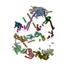
  • Deposited structure unit
  • Imaged by Jmol
  • Download
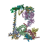
  • Simplified surface model + fitted atomic model
  • EMDB-2787
  • Imaged by Jmol
  • Download
Movie viewer
Structure viewerMolecule:
MolmilJmol/JSmol

Downloads & links

-
Assembly

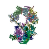
Deposited unit
a: MITORIBOSOMAL PROTEIN ML37, MRPL37
b: MITORIBOSOMAL PROTEIN ML38, MRPL38
c: MITORIBOSOMAL PROTEIN ML39, MRPL39
d: MITORIBOSOMAL PROTEIN ML40, MRPL40
e: MITORIBOSOMAL PROTEIN ML41, MRPL41
f: MITORIBOSOMAL PROTEIN ML42, MRPL42
g: MITORIBOSOMAL PROTEIN ML43, MRPL43
h: MITORIBOSOMAL PROTEIN ML44, MRPL44
i: MITORIBOSOMAL PROTEIN ML45, MRPL45
j: MITORIBOSOMAL PROTEIN ML46, MRPL46
k: MITORIBOSOMAL PROTEIN ML48, MRPL48
l: MITORIBOSOMAL PROTEIN ML49, MRPL49
m: MITORIBOSOMAL PROTEIN ML50, MRPL50
n: MITORIBOSOMAL PROTEIN ML51, MRPL51
o: MITORIBOSOMAL PROTEIN ML52, MRPL52
p: MITORIBOSOMAL PROTEIN ML53, MRPL53
q: MITORIBOSOMAL PROTEIN ML54, MRPL54
t: MITORIBOSOMAL PROTEIN ML63, MRPL57, MRP63
u: MITORIBOSOMAL PROTEIN ML62, MRPL58, ICT1
v: MITORIBOSOMAL PROTEIN ML64, MRPL59, CRIF1
w: MITORIBOSOMAL PROTEIN ML65, MRPS30
x: MITORIBOSOMAL PROTEIN ML66, MRPS18A
z: UNASSIGNED SECONDARY STRUCTURE ELEMENTS
hetero molecules


Theoretical massNumber of molelcules
Total (without water)563,88924
Polymers563,82323
Non-polymers651
Water00
1


  • Idetical with deposited unit
  • defined by author&software
TypeNameSymmetry operationNumber
identity operation1_555x,y,z1
MethodPISA

-
Components

+
MITORIBOSOMAL PROTEIN ... , 22 types, 22 molecules abcdefghijklmnopqtuvwx

#1: Protein MITORIBOSOMAL PROTEIN ML37, MRPL37


Mass: 47935.961 Da / Num. of mol.: 1 / Source method: isolated from a natural source / Source: (natural) SUS SCROFA (pig) / Organ: LIVER
#2: Protein MITORIBOSOMAL PROTEIN ML38, MRPL38


Mass: 44656.387 Da / Num. of mol.: 1 / Source method: isolated from a natural source / Source: (natural) SUS SCROFA (pig) / Organ: LIVER / References: UniProt: F1RW03
#3: Protein MITORIBOSOMAL PROTEIN ML39, MRPL39


Mass: 38523.422 Da / Num. of mol.: 1 / Source method: isolated from a natural source / Source: (natural) SUS SCROFA (pig) / Organ: LIVER / References: UniProt: F1SHP6*PLUS
#4: Protein MITORIBOSOMAL PROTEIN ML40, MRPL40


Mass: 24075.658 Da / Num. of mol.: 1 / Source method: isolated from a natural source / Source: (natural) SUS SCROFA (pig) / Organ: LIVER / References: UniProt: F1RK59
#5: Protein MITORIBOSOMAL PROTEIN ML41, MRPL41


Mass: 15073.404 Da / Num. of mol.: 1 / Source method: isolated from a natural source / Source: (natural) SUS SCROFA (pig) / Organ: LIVER
#6: Protein MITORIBOSOMAL PROTEIN ML42, MRPL42


Mass: 15908.536 Da / Num. of mol.: 1 / Source method: isolated from a natural source / Source: (natural) SUS SCROFA (pig) / Organ: LIVER
#7: Protein MITORIBOSOMAL PROTEIN ML43, MRPL43


Mass: 17728.109 Da / Num. of mol.: 1 / Source method: isolated from a natural source / Source: (natural) SUS SCROFA (pig) / Organ: LIVER / References: UniProt: F1S8U4
#8: Protein MITORIBOSOMAL PROTEIN ML44, MRPL44


Mass: 37527.195 Da / Num. of mol.: 1 / Source method: isolated from a natural source / Source: (natural) SUS SCROFA (pig) / Organ: LIVER / References: UniProt: F1SR64
#9: Protein MITORIBOSOMAL PROTEIN ML45, MRPL45


Mass: 36119.660 Da / Num. of mol.: 1 / Source method: isolated from a natural source / Source: (natural) SUS SCROFA (pig) / Organ: LIVER
#10: Protein MITORIBOSOMAL PROTEIN ML46, MRPL46


Mass: 32215.998 Da / Num. of mol.: 1 / Source method: isolated from a natural source / Source: (natural) SUS SCROFA (pig) / Organ: LIVER / References: UniProt: F1SRT0
#11: Protein MITORIBOSOMAL PROTEIN ML48, MRPL48


Mass: 23749.512 Da / Num. of mol.: 1 / Source method: isolated from a natural source / Source: (natural) SUS SCROFA (pig) / Organ: LIVER / References: UniProt: A0A480J9Z6*PLUS
#12: Protein MITORIBOSOMAL PROTEIN ML49, MRPL49


Mass: 18957.779 Da / Num. of mol.: 1 / Source method: isolated from a natural source / Source: (natural) SUS SCROFA (pig) / Organ: LIVER / References: UniProt: A0A0R4J8D5*PLUS
#13: Protein MITORIBOSOMAL PROTEIN ML50, MRPL50


Mass: 18351.000 Da / Num. of mol.: 1 / Source method: isolated from a natural source / Source: (natural) SUS SCROFA (pig) / Organ: LIVER / References: UniProt: I3L9H6
#14: Protein MITORIBOSOMAL PROTEIN ML51, MRPL51


Mass: 15219.983 Da / Num. of mol.: 1 / Source method: isolated from a natural source / Source: (natural) SUS SCROFA (pig) / Organ: LIVER
#15: Protein MITORIBOSOMAL PROTEIN ML52, MRPL52


Mass: 13813.705 Da / Num. of mol.: 1 / Source method: isolated from a natural source / Source: (natural) SUS SCROFA (pig) / Organ: LIVER / References: UniProt: F1S9B7
#16: Protein MITORIBOSOMAL PROTEIN ML53, MRPL53


Mass: 12015.747 Da / Num. of mol.: 1 / Source method: isolated from a natural source / Source: (natural) SUS SCROFA (pig) / Organ: LIVER / References: UniProt: F1SLH8
#17: Protein MITORIBOSOMAL PROTEIN ML54, MRPL54


Mass: 15957.357 Da / Num. of mol.: 1 / Source method: isolated from a natural source / Source: (natural) SUS SCROFA (pig) / Organ: LIVER
#18: Protein MITORIBOSOMAL PROTEIN ML63, MRPL57, MRP63


Mass: 12001.915 Da / Num. of mol.: 1 / Source method: isolated from a natural source / Source: (natural) SUS SCROFA (pig) / Organ: LIVER
#19: Protein MITORIBOSOMAL PROTEIN ML62, MRPL58, ICT1


Mass: 23281.758 Da / Num. of mol.: 1 / Source method: isolated from a natural source / Source: (natural) SUS SCROFA (pig) / Organ: LIVER / References: UniProt: W5IDC0
#20: Protein MITORIBOSOMAL PROTEIN ML64, MRPL59, CRIF1


Mass: 25347.646 Da / Num. of mol.: 1 / Source method: isolated from a natural source / Source: (natural) SUS SCROFA (pig) / Organ: LIVER / References: UniProt: F1SD95, PDB-4V1A95
#21: Protein MITORIBOSOMAL PROTEIN ML65, MRPS30


Mass: 49071.488 Da / Num. of mol.: 1 / Source method: isolated from a natural source / Source: (natural) SUS SCROFA (pig) / Organ: LIVER
#22: Protein MITORIBOSOMAL PROTEIN ML66, MRPS18A


Mass: 22273.084 Da / Num. of mol.: 1 / Source method: isolated from a natural source / Source: (natural) SUS SCROFA (pig) / Organ: LIVER / References: UniProt: F1RRH6

-
Protein/peptide / Non-polymers , 2 types, 2 molecules z

#23: Protein/peptide UNASSIGNED SECONDARY STRUCTURE ELEMENTS


Mass: 4017.944 Da / Num. of mol.: 1 / Source method: isolated from a natural source / Source: (natural) SUS SCROFA (pig) / Organ: LIVER
#24: Chemical ChemComp-ZN / ZINC ION


Mass: 65.409 Da / Num. of mol.: 1 / Source method: obtained synthetically / Formula: Zn

-
Details

Nonpolymer detailsUNKNOWN (UNK): UNASSIGNED AMINO ACIDS BUILT AS UNK ZINC ION (ZN): ZN IONS COORDINATED BY ZINC ...UNKNOWN (UNK): UNASSIGNED AMINO ACIDS BUILT AS UNK ZINC ION (ZN): ZN IONS COORDINATED BY ZINC FINGER PROTEINS CYS x70, CYS x73 AND CYS x108 FORM A MIXED ZINC FINGER TOGETHER WITH RESIDUE CYS J64 OF SPLIT ENTRY 4V19
Sequence detailsUNASSIGNED SECONDARY STRUCTURE ELEMENTS BUILT AS UNK RESIDUES OF v144-155 BUILT AS UNASSIGNED UNK ...UNASSIGNED SECONDARY STRUCTURE ELEMENTS BUILT AS UNK RESIDUES OF v144-155 BUILT AS UNASSIGNED UNK RESIDUES OF u164-173 BUILT AS UNASSIGNED UNK RESIDUES OF f77-100 BUILT AS UNASSIGNED UNK GENBANK REFERENCE FOR CHAIN a AK237653.1 GENBANK REFERENCE FOR CHAIN c XP_003132793.4 GENBANK REFERENCE FOR CHAIN e AW787117.1 GENBANK REFERENCE FOR CHAIN f AY609966.1 GENBANK REFERENCE FOR CHAIN i AK232067.1 GENBANK REFERENCE FOR CHAIN k AK391730.1 GENBANK REFERENCE FOR CHAIN l NP_001231942.1 GENBANK REFERENCE FOR CHAIN n EW306587.2 GENBANK REFERENCE FOR CHAIN q XP_003123104.1 GENBANK REFERENCE FOR CHAIN t AK347505.1 GENBANK REFERENCE FOR CHAIN w AK236026.1

-
Experimental details

-
Experiment

ExperimentMethod: ELECTRON MICROSCOPY
EM experimentAggregation state: PARTICLE / 3D reconstruction method: single particle reconstruction

-
Sample preparation

ComponentName: SUS SCROFA 55S MITOCHONDRIAL RIBOSOME / Type: RIBOSOME
Details: QUANTIFOIL HOLEY CARBON GRIDS WERE COATED WITH A THIN CARBON FILM
Buffer solutionName: 20 MM HEPES-KOH, 50 MM KCL, 40 MM MGCL2, 1 MM DTT / pH: 7.4 / Details: 20 MM HEPES-KOH, 50 MM KCL, 40 MM MGCL2, 1 MM DTT
SpecimenEmbedding applied: NO / Shadowing applied: NO / Staining applied: NO / Vitrification applied: YES
Specimen supportDetails: CARBON
VitrificationInstrument: HOMEMADE PLUNGER / Cryogen name: ETHANE-PROPANE / Details: MIXTURE OF LIQUID ETHANE AND PROPANE

-
Electron microscopy imaging

Experimental equipment
Model: Titan Krios / Image courtesy: FEI Company
MicroscopyModel: FEI TITAN KRIOS / Date: May 30, 2014
Details: IMAGES WERE ACQUIRED IN 2 SESSIONS ON A FEI TITAN KRIOS IN MAY 2014
Electron gunElectron source: FIELD EMISSION GUN / Accelerating voltage: 300 kV / Illumination mode: FLOOD BEAM
Electron lensMode: BRIGHT FIELD / Nominal magnification: 59000 X / Calibrated magnification: 100000 X / Nominal defocus max: 3000 nm / Nominal defocus min: 800 nm / Cs: 2.7 mm
Specimen holderTemperature: 85 K
Image recordingElectron dose: 20 e/Å2 / Film or detector model: FEI FALCON II (4k x 4k)
Radiation wavelengthRelative weight: 1

-
Processing

EM software
IDNameVersionCategory
1CTFFINDCTF correction
2Cootmodel fitting
3Omodel fitting
4UCSF Chimeramodel fitting
5EMAN1.93D reconstruction
6RELION3D reconstruction
CTF correctionDetails: PER DETECTOR FRAME
SymmetryPoint symmetry: C1 (asymmetric)
3D reconstructionMethod: MAXIMUM LIKELIHOOD BASED REFINEMENT IMPLEMENTED IN RELION
Resolution: 3.4 Å / Num. of particles: 141675 / Actual pixel size: 1.4 Å
Details: FOR VISUALIZATION PURPOSES THE FINAL MAP WAS FILTERED AND AMPLITUDE CORRECTED IN RELION SUBMISSION BASED ON EXPERIMENTAL DATA FROM EMDB EMD-2787. (DEPOSITION ID: 12830). THE COMBINED ...Details: FOR VISUALIZATION PURPOSES THE FINAL MAP WAS FILTERED AND AMPLITUDE CORRECTED IN RELION SUBMISSION BASED ON EXPERIMENTAL DATA FROM EMDB EMD-2787. (DEPOSITION ID: 12830). THE COMBINED COORDINATES (SPLIT ENTRIES 4V19 AND 4V1A) WERE REFINED IN RECIPROCAL SPACE USING PHENIX.REFINE AGAINST THE MLHL TARGET. FOR THIS, THE CRYO-EM MAPS (EMD-2787) WERE CONVERTED TO RECIPROCAL SPACE STRUCTURE FACTORS.
Symmetry type: POINT
Atomic model buildingSpace: REAL
RefinementHighest resolution: 3.4 Å
Refinement stepCycle: LAST / Highest resolution: 3.4 Å
ProteinNucleic acidLigandSolventTotal
Num. atoms31916 0 1 0 31917

+
About Yorodumi

-
News

-
Feb 9, 2022. New format data for meta-information of EMDB entries

New format data for meta-information of EMDB entries

  • Version 3 of the EMDB header file is now the official format.
  • The previous official version 1.9 will be removed from the archive.

Related info.:EMDB header

External links:wwPDB to switch to version 3 of the EMDB data model

-
Aug 12, 2020. Covid-19 info

Covid-19 info

URL: https://pdbj.org/emnavi/covid19.php

New page: Covid-19 featured information page in EM Navigator.

Related info.:Covid-19 info / Mar 5, 2020. Novel coronavirus structure data

+
Mar 5, 2020. Novel coronavirus structure data

Novel coronavirus structure data

Related info.:Yorodumi Speices / Aug 12, 2020. Covid-19 info

External links:COVID-19 featured content - PDBj / Molecule of the Month (242):Coronavirus Proteases

+
Jan 31, 2019. EMDB accession codes are about to change! (news from PDBe EMDB page)

EMDB accession codes are about to change! (news from PDBe EMDB page)

  • The allocation of 4 digits for EMDB accession codes will soon come to an end. Whilst these codes will remain in use, new EMDB accession codes will include an additional digit and will expand incrementally as the available range of codes is exhausted. The current 4-digit format prefixed with “EMD-” (i.e. EMD-XXXX) will advance to a 5-digit format (i.e. EMD-XXXXX), and so on. It is currently estimated that the 4-digit codes will be depleted around Spring 2019, at which point the 5-digit format will come into force.
  • The EM Navigator/Yorodumi systems omit the EMD- prefix.

Related info.:Q: What is EMD? / ID/Accession-code notation in Yorodumi/EM Navigator

External links:EMDB Accession Codes are Changing Soon! / Contact to PDBj

+
Jul 12, 2017. Major update of PDB

Major update of PDB

  • wwPDB released updated PDB data conforming to the new PDBx/mmCIF dictionary.
  • This is a major update changing the version number from 4 to 5, and with Remediation, in which all the entries are updated.
  • In this update, many items about electron microscopy experimental information are reorganized (e.g. em_software).
  • Now, EM Navigator and Yorodumi are based on the updated data.

External links:wwPDB Remediation / Enriched Model Files Conforming to OneDep Data Standards Now Available in the PDB FTP Archive

-
Yorodumi

Thousand views of thousand structures

  • Yorodumi is a browser for structure data from EMDB, PDB, SASBDB, etc.
  • This page is also the successor to EM Navigator detail page, and also detail information page/front-end page for Omokage search.
  • The word "yorodu" (or yorozu) is an old Japanese word meaning "ten thousand". "mi" (miru) is to see.

Related info.:EMDB / PDB / SASBDB / Comparison of 3 databanks / Yorodumi Search / Aug 31, 2016. New EM Navigator & Yorodumi / Yorodumi Papers / Jmol/JSmol / Function and homology information / Changes in new EM Navigator and Yorodumi

Read more