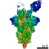
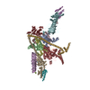
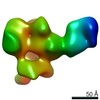
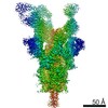
- PDB-3j9c: CryoEM single particle reconstruction of anthrax toxin protective... -
+
データを開く
IDまたはキーワード:
読み込み中...
-
基本情報
登録情報
データベース: PDB / ID: 3j9c
タイトル
CryoEM single particle reconstruction of anthrax toxin protective antigen pore at 2.9 Angstrom resolution
要素
Protective antigen PA-63
キーワード
TOXIN / TRANSPORT PROTEIN / bacterial toxin / anthrax toxin / protective antigen / protein translocation channel
機能・相同性
機能・相同性情報
positive regulation of apoptotic process in another organism / host cell cytosol / negative regulation of MAPK cascade / Uptake and function of anthrax toxins / host cell endosome membrane / protein homooligomerization / toxin activity / host cell plasma membrane / extracellular region / identical protein binding ...positive regulation of apoptotic process in another organism / host cell cytosol / negative regulation of MAPK cascade / Uptake and function of anthrax toxins / host cell endosome membrane / protein homooligomerization / toxin activity / host cell plasma membrane / extracellular region / identical protein binding / membrane / metal ion binding 類似検索 - 分子機能
ジャーナル: Nature / 年: 2015 タイトル: Atomic structure of anthrax protective antigen pore elucidates toxin translocation. 著者: Jiansen Jiang / Bradley L Pentelute / R John Collier / Z Hong Zhou / 要旨: Anthrax toxin, comprising protective antigen, lethal factor, and oedema factor, is the major virulence factor of Bacillus anthracis, an agent that causes high mortality in humans and animals. ...Anthrax toxin, comprising protective antigen, lethal factor, and oedema factor, is the major virulence factor of Bacillus anthracis, an agent that causes high mortality in humans and animals. Protective antigen forms oligomeric prepores that undergo conversion to membrane-spanning pores by endosomal acidification, and these pores translocate the enzymes lethal factor and oedema factor into the cytosol of target cells. Protective antigen is not only a vaccine component and therapeutic target for anthrax infections but also an excellent model system for understanding the mechanism of protein translocation. On the basis of biochemical and electrophysiological results, researchers have proposed that a phi (Φ)-clamp composed of phenylalanine (Phe)427 residues of protective antigen catalyses protein translocation via a charge-state-dependent Brownian ratchet. Although atomic structures of protective antigen prepores are available, how protective antigen senses low pH, converts to active pore, and translocates lethal factor and oedema factor are not well defined without an atomic model of its pore. Here, by cryo-electron microscopy with direct electron counting, we determine the protective antigen pore structure at 2.9-Å resolution. The structure reveals the long-sought-after catalytic Φ-clamp and the membrane-spanning translocation channel, and supports the Brownian ratchet model for protein translocation. Comparisons of four structures reveal conformational changes in prepore to pore conversion that support a multi-step mechanism by which low pH is sensed and the membrane-spanning channel is formed.
 ムービー
ムービー コントローラー


 データを開く
 基本情報
基本情報 要素
要素 キーワード
キーワード 機能・相同性情報
 データ登録者
 引用
引用
 構造の表示
構造の表示 ダウンロードとリンク
 その他のダウンロード
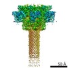


 PDBj
PDBj


 集合体
集合体


 分子量: 40.078 Da / 分子数: 2 / 由来タイプ: 合成 / 式: Ca
 試料調製
試料調製 電子顕微鏡撮影
 FIELD EMISSION GUN / 加速電圧: 300 kV / 照射モード: FLOOD BEAM
 解析
解析