 Movie
Movie Controller


+ Open data

- Basic information
| Entry | Database: PDB / ID: 3gzu | ||||||
|---|---|---|---|---|---|---|---|
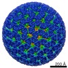
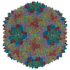
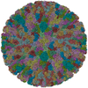
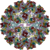
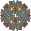
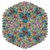
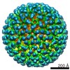
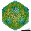
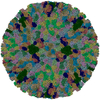
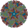
| Title | VP7 recoated rotavirus DLP | ||||||
 Components Components |
| ||||||
 Keywords Keywords | VIRUS / rotavirus / VP7 / VP6 / VP2 / 7RP / DLP / Capsid protein / Metal-binding / Virion / Zinc / Core protein / RNA-binding / Icosaderal virus | ||||||
| Function / homology |  Function and homology information Function and homology informationviral intermediate capsid / T=2 icosahedral viral capsid / T=13 icosahedral viral capsid / viral inner capsid / viral nucleocapsid / host cell surface receptor binding / fusion of virus membrane with host plasma membrane / viral envelope / structural molecule activity / RNA binding / metal ion binding Similarity search - Function | ||||||
| Biological species |  Rotavirus ARhesus Rotavirus Rotavirus ARhesus Rotavirus | ||||||
| Method | ELECTRON MICROSCOPY / single particle reconstruction / cryo EM / Resolution: 3.8 Å | ||||||
 Authors Authors | Chen, J.Z. / Settembre, E.C. / Harrison, S.C. / Grigorieff, N. | ||||||
 Citation Citation | Journal: Proc Natl Acad Sci U S A / Year: 2009 Title: Molecular interactions in rotavirus assembly and uncoating seen by high-resolution cryo-EM. Authors: James Z Chen / Ethan C Settembre / Scott T Aoki / Xing Zhang / A Richard Bellamy / Philip R Dormitzer / Stephen C Harrison / Nikolaus Grigorieff /  Abstract: Rotaviruses, major causes of childhood gastroenteritis, are nonenveloped, icosahedral particles with double-strand RNA genomes. By the use of electron cryomicroscopy and single-particle ...Rotaviruses, major causes of childhood gastroenteritis, are nonenveloped, icosahedral particles with double-strand RNA genomes. By the use of electron cryomicroscopy and single-particle reconstruction, we have visualized a rotavirus particle comprising the inner capsid coated with the trimeric outer-layer protein, VP7, at a resolution (4 A) comparable with that of X-ray crystallography. We have traced the VP7 polypeptide chain, including parts not seen in its X-ray crystal structure. The 3 well-ordered, 30-residue, N-terminal "arms" of each VP7 trimer grip the underlying trimer of VP6, an inner-capsid protein. Structural differences between free and particle-bound VP7 and between free and VP7-coated inner capsids may regulate mRNA transcription and release. The Ca(2+)-stabilized VP7 intratrimer contact region, which presents important neutralizing epitopes, is unaltered upon capsid binding. | ||||||
| History |
|
- Structure visualization
| Movie |
Movie viewer |
|---|---|
| Structure viewer | Molecule: MolmilJmol/JSmol |
- Downloads & links
-Download
| PDBx/mmCIF format | 3gzu.cif.gz | 1.2 MB | Display | PDBx/mmCIF format |
|---|---|---|---|---|
| PDB format | pdb3gzu.ent.gz | 994.9 KB | Display | PDB format |
| PDBx/mmJSON format | 3gzu.json.gz | Tree view | PDBx/mmJSON format | |
| Others |  Other downloads Other downloads |
-Validation report
| Summary document | 3gzu_validation.pdf.gz | 930.4 KB | Display | wwPDB validaton report |
|---|---|---|---|---|
| Full document | 3gzu_full_validation.pdf.gz | 1.3 MB | Display | |
| Data in XML | 3gzu_validation.xml.gz | 218.2 KB | Display | |
| Data in CIF | 3gzu_validation.cif.gz | 319.4 KB | Display | |
| Arichive directory | https://data.pdbj.org/pub/pdb/validation_reports/gz/3gzuftp://data.pdbj.org/pub/pdb/validation_reports/gz/3gzu | HTTPS FTP |
-Related structure data


-Links
 PDBj
PDBj


- Assembly
| Deposited unit | 
|
|---|---|
| 1 | x 60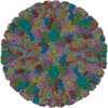
|
| 2 |
|
| 3 | x 5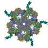
|
| 4 | x 6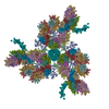
|
| 5 | 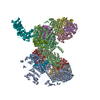
|
| Symmetry | Point symmetry: (Schoenflies symbol: I (icosahedral)) |
-Components
| #1: Protein | Mass: 93127.438 Da / Num. of mol.: 2 / Fragment: VP2 Source method: isolated from a genetically manipulated source Source: (gene. exp.) Rotavirus A / Production host:   Spodoptera frugiperda (fall armyworm) / References: UniProt: B2BMF8, UniProt: B2BMD1*PLUS Spodoptera frugiperda (fall armyworm) / References: UniProt: B2BMF8, UniProt: B2BMD1*PLUS#2: Protein | Mass: 44879.641 Da / Num. of mol.: 13 / Fragment: VP6 Source method: isolated from a genetically manipulated source Source: (gene. exp.) Rhesus Rotavirus / Production host: Spodoptera frugiperda (fall armyworm) / Strain (production host): Sf9 / References: UniProt: P04509#3: Chemical | ChemComp-ZN /   Mass: 65.409 Da / Num. of mol.: 5 / Source method: obtained synthetically / Formula: Zn Mass: 65.409 Da / Num. of mol.: 5 / Source method: obtained synthetically / Formula: Zn |
|---|
-Experimental details
-Experiment
| Experiment | Method: ELECTRON MICROSCOPY |
|---|---|
| EM experiment | Aggregation state: PARTICLE / 3D reconstruction method: single particle reconstruction |
- Sample preparation
| Component | Name: VP7 recoated rotavirus DLP / Type: VIRUS / Details: capsid protein VP7. VP6 and VP2 |
|---|---|
| Details of virus | Host category: REOVIRIDAE / Type: VIRUS-LIKE PARTICLE |
| Natural host | Organism: Bos taurus |
| Buffer solution | Name: 20mM TrisHCl, 50mM NaCl, 2mM CaCl2 / pH: 8 / Details: 20mM TrisHCl, 50mM NaCl, 2mM CaCl2 |
| Specimen | Conc.: 5 mg/ml / Embedding applied: NO / Shadowing applied: NO / Staining applied: NO / Vitrification applied: YES |
| Specimen support | Details: C-flat grids |
| Vitrification | Instrument: HOMEMADE PLUNGER / Cryogen name: ETHANE / Details: manual plunging at 90K |
- Electron microscopy imaging
| Experimental equipment |  Model: Tecnai F30 / Image courtesy: FEI Company |
|---|---|
| Microscopy | Model: FEI TECNAI F30 / Date: Dec 1, 2007 |
| Electron gun | Electron source:  FIELD EMISSION GUN / Accelerating voltage: 300 kV / Illumination mode: FLOOD BEAM FIELD EMISSION GUN / Accelerating voltage: 300 kV / Illumination mode: FLOOD BEAM |
| Electron lens | Mode: BRIGHT FIELD / Nominal magnification: 59000 X / Calibrated magnification: 58168 X / Nominal defocus max: 3500 nm / Nominal defocus min: 1200 nm / Cs: 2 mm |
| Specimen holder | Temperature: 90 K / Tilt angle max: 0 ° / Tilt angle min: 0 ° |
| Image recording | Electron dose: 25 e/Å2 / Film or detector model: GENERIC FILM / Details: Kodak ISO163 |
- Processing
| CTF correction | Details: individual particle CTF | ||||||||||||
|---|---|---|---|---|---|---|---|---|---|---|---|---|---|
| Symmetry | Point symmetry: I (icosahedral) | ||||||||||||
| 3D reconstruction | Method: projection matching and refinement using FEALIGN / Resolution: 3.8 Å / Num. of particles: 3780 / Nominal pixel size: 1.233 Å / Actual pixel size: 1.233 Å / Magnification calibration: 58168 / Details: projection matching by FREALIGN / Symmetry type: POINT | ||||||||||||
| Atomic model building | Space: REAL | ||||||||||||
| Refinement step | Cycle: LAST
|
