 Movie
Movie Controller

[English] 日本語
 Yorodumi
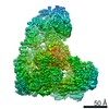
- PDB-3a69: Atomic model of the bacterial flagellar hook based on docking an ... -
+ Open data

- Basic information
| Entry | Database: PDB / ID: 3a69 | ||||||
|---|---|---|---|---|---|---|---|
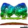
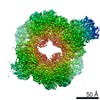
| Title | Atomic model of the bacterial flagellar hook based on docking an X-ray derived structure and terminal two alpha-helices into an 7.1 angstrom resolution cryoEM map | ||||||
 Components Components | Flagellar hook protein flgE | ||||||
 Keywords Keywords | MOTOR PROTEIN / the bacterial flagellar motor / universal joint / Bacterial flagellum | ||||||
| Function / homology |  Function and homology information Function and homology informationbacterial-type flagellum basal body / bacterial-type flagellum-dependent swarming motility Similarity search - Function | ||||||
| Biological species |  Salmonella enterica subsp. enterica serovar Typhimurium (bacteria) Salmonella enterica subsp. enterica serovar Typhimurium (bacteria) | ||||||
| Method | ELECTRON MICROSCOPY / helical reconstruction / cryo EM / Resolution: 7.1 Å | ||||||
 Authors Authors | Fujii, T. / Kato, T. / Namba, K. | ||||||
 Citation Citation | Journal: Structure / Year: 2009 Title: Specific arrangement of alpha-helical coiled coils in the core domain of the bacterial flagellar hook for the universal joint function. Authors: Takashi Fujii / Takayuki Kato / Keiichi Namba /  Abstract: The bacterial flagellar hook is a short, highly curved tubular structure connecting the rotary motor to the filament acting as a helical propeller. The bending flexibility of the hook allows it to ...The bacterial flagellar hook is a short, highly curved tubular structure connecting the rotary motor to the filament acting as a helical propeller. The bending flexibility of the hook allows it to work as a universal joint. A partial atomic model of the hook revealed a sliding intersubunit domain interaction along the protofilament to produce bending flexibility. However, it remained unclear how the tightly packed inner core domains can still permit axial extension and compression. We report advances in cryoEM image analysis for high-resolution, high-throughput structural analysis and a density map of the hook that reveals most of the secondary structures, including the terminal alpha helices forming a coiled coil. The orientations and axial packing interactions of these two alpha helices are distinctly different from those of the filament, allowing them to have a room for axial compression and extension for bending flexibility without impairing the mechanical stability of the hook. | ||||||
| History |
|
- Structure visualization
| Movie |
Movie viewer |
|---|---|
| Structure viewer | Molecule: MolmilJmol/JSmol |
- Downloads & links
-Download
| PDBx/mmCIF format | 3a69.cif.gz | 72.9 KB | Display | PDBx/mmCIF format |
|---|---|---|---|---|
| PDB format | pdb3a69.ent.gz | 53.9 KB | Display | PDB format |
| PDBx/mmJSON format | 3a69.json.gz | Tree view | PDBx/mmJSON format | |
| Others |  Other downloads Other downloads |
-Validation report
| Summary document | 3a69_validation.pdf.gz | 809.6 KB | Display | wwPDB validaton report |
|---|---|---|---|---|
| Full document | 3a69_full_validation.pdf.gz | 817.9 KB | Display | |
| Data in XML | 3a69_validation.xml.gz | 18.1 KB | Display | |
| Data in CIF | 3a69_validation.cif.gz | 24.8 KB | Display | |
| Arichive directory | https://data.pdbj.org/pub/pdb/validation_reports/a6/3a69ftp://data.pdbj.org/pub/pdb/validation_reports/a6/3a69 | HTTPS FTP |
-Related structure data
| Related structure data | 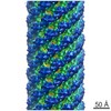 1647MC M: map data used to model this data C: citing same article ( |
|---|---|
| Similar structure data |

-Links
 PDBj
PDBj
- Assembly
| Deposited unit | 
|
|---|---|
| 1 | x 11
|
| 2 |
|
| 3 | 
|
| Symmetry | Helical symmetry: (Circular symmetry: 1 / Dyad axis: no / N subunits divisor: 1 / Num. of operations: 11 / Rise per n subunits: 4.123 Å / Rotation per n subunits: 64.786 °) |
-Components
| #1: Protein | Mass: 42101.957 Da / Num. of mol.: 1 / Source method: isolated from a natural source Source: (natural) Salmonella enterica subsp. enterica serovar Typhimurium (bacteria)Strain: SJW880 / References: UniProt: P0A1J1 |
|---|
-Experimental details
-Experiment
| Experiment | Method: ELECTRON MICROSCOPY |
|---|---|
| EM experiment | Aggregation state: FILAMENT / 3D reconstruction method: helical reconstruction |
- Sample preparation
| Component | Name: bacterial flagellar hook / Type: COMPLEX |
|---|---|
| Buffer solution | Name: 20mM Tris-HCl, 100mM NaCl / pH: 7 / Details: 20mM Tris-HCl, 100mM NaCl |
| Specimen | Embedding applied: NO / Shadowing applied: NO / Staining applied: NO / Vitrification applied: YES |
| Vitrification | Instrument: FEI VITROBOT MARK I / Cryogen name: HELIUM / Humidity: 90 % |
- Electron microscopy imaging
| Microscopy | Model: JEOL 3200FSC / Date: Feb 20, 2008 |
|---|---|
| Electron gun | Electron source:  FIELD EMISSION GUN / Accelerating voltage: 200 kV / Illumination mode: FLOOD BEAM FIELD EMISSION GUN / Accelerating voltage: 200 kV / Illumination mode: FLOOD BEAM |
| Electron lens | Mode: BRIGHT FIELD / Nominal magnification: 50000 X / Calibrated magnification: 89285 X / Nominal defocus max: 2000 nm / Nominal defocus min: 500 nm / Cs: 1.6 mm |
| Specimen holder | Specimen holder model: JEOL Specimen holder type: Top entry liquid helium-cooled cryo specimen holder Temperature: 50 K |
| Image recording | Electron dose: 20 e/Å2 / Film or detector model: TVIPS TEMCAM-F415 (4k x 4k) |
| EM imaging optics | Energyfilter upper: 10 eV / Energyfilter lower: 0 eV |
| Radiation | Protocol: SINGLE WAVELENGTH / Monochromatic (M) / Laue (L): M |
| Radiation wavelength | Relative weight: 1 |
- Processing
| CTF correction | Details: each images | ||||||||||||
|---|---|---|---|---|---|---|---|---|---|---|---|---|---|
| 3D reconstruction | Method: IHRSR / Resolution: 7.1 Å / Actual pixel size: 1.68 Å / Magnification calibration: TMV images Details: a modified version of SPIDER program was used for the reconstruction Symmetry type: HELICAL | ||||||||||||
| Refinement step | Cycle: LAST
|
