 Movie
Movie Controller

[English] 日本語
 Yorodumi
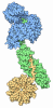
- PDB-35zv: yeast 26S proteasome base assembly intermediate, Rpn14-Rpt6 (base... -
+ Open data

- Basic information
| Entry | Database: PDB / ID: 35zv | |||||||||
|---|---|---|---|---|---|---|---|---|---|---|
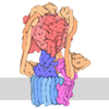
| Title | yeast 26S proteasome base assembly intermediate, Rpn14-Rpt6 (base-Rpn14-Hsm3-Nas6(2) with Rpt4-EQ) | |||||||||
 Components Components |
| |||||||||
 Keywords Keywords | MOTOR PROTEIN / proteasome / chaperone / base / 26S / AAA / motor / assembly | |||||||||
| Function / homology |  Function and homology information Function and homology informationproteasome regulatory particle, base subcomplex / proteasome regulatory particle assembly / proteasome-activating activity / regulation of protein catabolic process / Cross-presentation of soluble exogenous antigens (endosomes) / TNFR2 non-canonical NF-kB pathway / Proteasome assembly / nonfunctional rRNA decay / Ub-specific processing proteases / positive regulation of RNA polymerase II transcription preinitiation complex assembly ...proteasome regulatory particle, base subcomplex / proteasome regulatory particle assembly / proteasome-activating activity / regulation of protein catabolic process / Cross-presentation of soluble exogenous antigens (endosomes) / TNFR2 non-canonical NF-kB pathway / Proteasome assembly / nonfunctional rRNA decay / Ub-specific processing proteases / positive regulation of RNA polymerase II transcription preinitiation complex assembly / enzyme regulator activity / Neutrophil degranulation / proteasome storage granule / protein folding chaperone / proteasome complex / positive regulation of transcription elongation by RNA polymerase II / nucleotide-excision repair / ubiquitin-dependent protein catabolic process / proteasome-mediated ubiquitin-dependent protein catabolic process / protein-macromolecule adaptor activity / chromatin remodeling / protein domain specific binding / ubiquitin protein ligase binding / endoplasmic reticulum / ATP hydrolysis activity / ATP binding / identical protein binding / nucleus / cytosol / cytoplasm Similarity search - Function | |||||||||
| Biological species |   Saccharomyces cerevisiae (brewer's yeast) Saccharomyces cerevisiae (brewer's yeast) | |||||||||
| Method | ELECTRON MICROSCOPY / single particle reconstruction / cryo EM / Resolution: 3.11 Å | |||||||||
 Authors Authors | Hsieh, H.H. / Martin, A. | |||||||||
| Funding support |  United States, 2items United States, 2items
| |||||||||
 Citation Citation | Journal: bioRxiv / Year: 2026 Title: Chaperones shape the conformational landscape of 26S-proteasome-base assembly for allosteric ATPase motor activation. Authors: Hao-Hsuan Hsieh / Andreas Martin / Abstract: Protein homeostasis depends on the 26S proteasome, the most complex ATP-dependent protease in eukaryotic cells. The proteasome base subcomplex is responsible for mechanical substrate unfolding and ...Protein homeostasis depends on the 26S proteasome, the most complex ATP-dependent protease in eukaryotic cells. The proteasome base subcomplex is responsible for mechanical substrate unfolding and translocation into an internal degradation chamber. It contains three non-ATPase subunits, Rpn1, Rpn2, and Rpn13, and a heterohexameric AAA+ motor with six distinct ATPases, Rpt1 - Rpt6. Correct base assembly requires four dedicated chaperones that initially form the Hsm3 module (Hsm3-Rpt1-Rpt2-Rpn1), the Rpn14/Nas6 module (Rpn14-Rpt6-Nas6-Rpt3-Rpn2-Rpn13), and the Nas2 module (Nas2-Rpt5-Rpt4). However, the mechanisms underlying module assembly and formation of the mature base remain unknown. Here, we reconstitute the base subcomplex of the 26S proteasome from recombinant modules. Using biochemical assays, mass photometry, single-molecule fluorescence measurements, and single-particle cryo-EM, we reveal how the chaperones direct the conformational transitions through several intermediates toward the ATP-hydrolysis-active base. The Nas2 and Rpn14/Nas6 modules associate first, and binding of the Hsm3 module creates a state in which the chaperones stabilize an open ATPase ring that lacks hydrolysis activity. Sequential chaperone release then leads to a gradual ATPase-ring closure, whereby Hsm3's unstructured C-terminal tail mimics a substrate polypeptide in the central channel and induces a processing motor state with a spiral-staircase arrangement of Rpt subunits and a closed ATPase site at Rpt4. Inaugural ATP hydrolysis in Rpt4 is subsequently required to eject Hsm3 and transition to the Nas6-bound base that is ATPase active and competent for 26S-proteasome incorporation. Our studies thus provide exciting insights into how chaperones assure correct assembly, guide the complex through an intricate conformational landscape, and thereby prevent premature ATP-hydrolysis activation or incorporation of faulty assemblies into holoenzymes. #1: Journal: Acta Crystallogr D Struct Biol / Year: 2019 Title: Macromolecular structure determination using X-rays, neutrons and electrons: recent developments in Phenix. Authors: Dorothee Liebschner / Pavel V Afonine / Matthew L Baker / Gábor Bunkóczi / Vincent B Chen / Tristan I Croll / Bradley Hintze / Li Wei Hung / Swati Jain / Airlie J McCoy / Nigel W Moriarty ...Authors: Dorothee Liebschner / Pavel V Afonine / Matthew L Baker / Gábor Bunkóczi / Vincent B Chen / Tristan I Croll / Bradley Hintze / Li Wei Hung / Swati Jain / Airlie J McCoy / Nigel W Moriarty / Robert D Oeffner / Billy K Poon / Michael G Prisant / Randy J Read / Jane S Richardson / David C Richardson / Massimo D Sammito / Oleg V Sobolev / Duncan H Stockwell / Thomas C Terwilliger / Alexandre G Urzhumtsev / Lizbeth L Videau / Christopher J Williams / Paul D Adams /   Abstract: Diffraction (X-ray, neutron and electron) and electron cryo-microscopy are powerful methods to determine three-dimensional macromolecular structures, which are required to understand biological ...Diffraction (X-ray, neutron and electron) and electron cryo-microscopy are powerful methods to determine three-dimensional macromolecular structures, which are required to understand biological processes and to develop new therapeutics against diseases. The overall structure-solution workflow is similar for these techniques, but nuances exist because the properties of the reduced experimental data are different. Software tools for structure determination should therefore be tailored for each method. Phenix is a comprehensive software package for macromolecular structure determination that handles data from any of these techniques. Tasks performed with Phenix include data-quality assessment, map improvement, model building, the validation/rebuilding/refinement cycle and deposition. Each tool caters to the type of experimental data. The design of Phenix emphasizes the automation of procedures, where possible, to minimize repetitive and time-consuming manual tasks, while default parameters are chosen to encourage best practice. A graphical user interface provides access to many command-line features of Phenix and streamlines the transition between programs, project tracking and re-running of previous tasks. | |||||||||
| History |
|
- Structure visualization
| Structure viewer | Molecule: MolmilJmol/JSmol |
|---|
- Downloads & links
-Download
| PDBx/mmCIF format | 35zv.cif.gz | 239.7 KB | Display | PDBx/mmCIF format |
|---|---|---|---|---|
| PDB format | pdb35zv.ent.gz | 139.1 KB | Display | PDB format |
| PDBx/mmJSON format | 35zv.json.gz | Tree view | PDBx/mmJSON format | |
| Others |  Other downloads Other downloads |
-Validation report
| Arichive directory | https://data.pdbj.org/pub/pdb/validation_reports/5z/35zvftp://data.pdbj.org/pub/pdb/validation_reports/5z/35zv | HTTPS FTP |
|---|
-Related structure data
| Related structure data |  77307MC  35zrC  35zwC  36axC  36bdC  36blC  36bmC C: citing same article ( M: map data used to model this data |
|---|---|
| Similar structure data |


-Links
 PDBj
PDBj


- Assembly
| Deposited unit | 
|
|---|---|
| 1 |
|
-Components
| #1: Protein | Mass: 109601.906 Da / Num. of mol.: 1 Source method: isolated from a genetically manipulated source Source: (gene. exp.) Saccharomyces cerevisiae (brewer's yeast)Gene: RPN1, HRD2, NAS1, RPD1, YHR027C / Production host:   Escherichia coli (E. coli) / References: UniProt: P38764 Escherichia coli (E. coli) / References: UniProt: P38764 |
|---|---|
| #2: Protein | Mass: 45342.742 Da / Num. of mol.: 1 Source method: isolated from a genetically manipulated source Source: (gene. exp.) Saccharomyces cerevisiae (brewer's yeast)Gene: RPT6, CIM3, CRL3, SUG1, TBPY, TBY1, YGL048C / Production host: Escherichia coli (E. coli) / References: UniProt: Q01939 |
| #3: Protein | Mass: 47953.676 Da / Num. of mol.: 1 Source method: isolated from a genetically manipulated source Source: (gene. exp.) Saccharomyces cerevisiae (brewer's yeast)Gene: RPT3, YNT1, YTA2, YDR394W, D9509.14 / Production host: Escherichia coli (E. coli) / References: UniProt: P33298 |
| #4: Protein | Mass: 46433.684 Da / Num. of mol.: 1 Source method: isolated from a genetically manipulated source Source: (gene. exp.) Saccharomyces cerevisiae (brewer's yeast)Gene: RPN14, YGL004C / Production host: Escherichia coli (E. coli) / References: UniProt: P53196 |
| #5: Chemical | ChemComp-ATP / 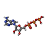 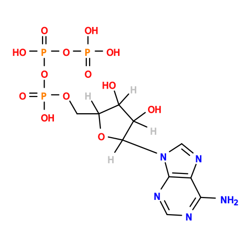 Mass: 507.181 Da / Num. of mol.: 1 / Source method: obtained synthetically / Formula: C10H16N5O13P3 / Feature type: SUBJECT OF INVESTIGATION / Comment: ATP, energy-carrying molecule*YM Mass: 507.181 Da / Num. of mol.: 1 / Source method: obtained synthetically / Formula: C10H16N5O13P3 / Feature type: SUBJECT OF INVESTIGATION / Comment: ATP, energy-carrying molecule*YM |
| Has ligand of interest | Y |
| Has protein modification | N |
-Experimental details
-Experiment
| Experiment | Method: ELECTRON MICROSCOPY |
|---|---|
| EM experiment | Aggregation state: PARTICLE / 3D reconstruction method: single particle reconstruction |
- Sample preparation
| Component | Name: yeast 26S proteasome base assembly intermediate, Hsm3-Rpt1-Rpt2 (base-Hsm3-Nas6) Type: COMPLEX / Entity ID: #1-#4 / Source: RECOMBINANT |
|---|---|
| Source (natural) | Organism: Saccharomyces cerevisiae (brewer's yeast) |
| Source (recombinant) | Organism: Escherichia coli (E. coli) |
| Buffer solution | pH: 7.6 |
| Specimen | Conc.: 6 mg/ml / Embedding applied: NO / Shadowing applied: NO / Staining applied: NO / Vitrification applied: YES |
| Vitrification | Cryogen name: ETHANE |
- Electron microscopy imaging
| Experimental equipment |  Model: Titan Krios / Image courtesy: FEI Company |
|---|---|
| Microscopy | Model: TFS KRIOS |
| Electron gun | Electron source:  FIELD EMISSION GUN / Accelerating voltage: 300 kV / Illumination mode: FLOOD BEAM FIELD EMISSION GUN / Accelerating voltage: 300 kV / Illumination mode: FLOOD BEAM |
| Electron lens | Mode: BRIGHT FIELD / Nominal defocus max: 2000 nm / Nominal defocus min: 500 nm |
| Image recording | Electron dose: 50 e/Å2 / Film or detector model: GATAN K3 (6k x 4k) |
- Processing
| EM software |
| ||||||||||||||||||||||||
|---|---|---|---|---|---|---|---|---|---|---|---|---|---|---|---|---|---|---|---|---|---|---|---|---|---|
| CTF correction | Type: PHASE FLIPPING AND AMPLITUDE CORRECTION | ||||||||||||||||||||||||
| 3D reconstruction | Resolution: 3.11 Å / Resolution method: FSC 0.143 CUT-OFF / Num. of particles: 208110 / Symmetry type: POINT | ||||||||||||||||||||||||
| Refinement | Cross valid method: NONE Stereochemistry target values: GeoStd + Monomer Library + CDL v1.2 | ||||||||||||||||||||||||
| Displacement parameters | Biso mean: 102.07 Å2 | ||||||||||||||||||||||||
| Refine LS restraints |
|
