 ムービー
ムービー コントローラー


+ データを開く

- 基本情報
基本情報
| 登録情報 | データベース: PDB / ID: 2ypw | ||||||
|---|---|---|---|---|---|---|---|
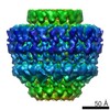
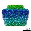
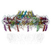
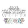
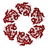
| タイトル | Atomic model for the N-terminus of TraO fitted in the full-length structure of the bacterial pKM101 type IV secretion system core complex | ||||||
 要素 要素 | TRAO | ||||||
 キーワード キーワード | MEMBRANE PROTEIN / BACTERIAL SECRETION / TYPE IV SECRETION | ||||||
| 機能・相同性 | Conjugal transfer, TrbG/VirB9/CagX / VirB9/CagX/TrbG, C-terminal / VirB9/CagX/TrbG, C-terminal domain superfamily / Conjugal transfer protein / TraO protein 機能・相同性情報 機能・相同性情報 | ||||||
| 生物種 |  | ||||||
| 手法 | 電子顕微鏡法 / 単粒子再構成法 / クライオ電子顕微鏡法 / 解像度: 12.4 Å | ||||||
 データ登録者 データ登録者 | Rivera-Calzada, A. / Fronzes, R. / Savva, C.G. / Chandran, V. / Lian, P.W. / Laeremans, T. / Pardon, E. / Steyaert, J. / Remaut, H. / Waksman, G. / Orlova, E.V. | ||||||
 引用 引用 | ジャーナル: EMBO J / 年: 2013 タイトル: Structure of a bacterial type IV secretion core complex at subnanometre resolution. 著者: Angel Rivera-Calzada / Rémi Fronzes / Christos G Savva / Vidya Chandran / Pei W Lian / Toon Laeremans / Els Pardon / Jan Steyaert / Han Remaut / Gabriel Waksman / Elena V Orlova /  要旨: Type IV secretion (T4S) systems are able to transport DNAs and/or proteins through the membranes of bacteria. They form large multiprotein complexes consisting of 12 proteins termed VirB1-11 and ...Type IV secretion (T4S) systems are able to transport DNAs and/or proteins through the membranes of bacteria. They form large multiprotein complexes consisting of 12 proteins termed VirB1-11 and VirD4. VirB7, 9 and 10 assemble into a 1.07 MegaDalton membrane-spanning core complex (CC), around which all other components assemble. This complex is made of two parts, the O-layer inserted in the outer membrane and the I-layer inserted in the inner membrane. While the structure of the O-layer has been solved by X-ray crystallography, there is no detailed structural information on the I-layer. Using high-resolution cryo-electron microscopy and molecular modelling combined with biochemical approaches, we determined the I-layer structure and located its various components in the electron density. Our results provide new structural insights on the CC, from which the essential features of T4S system mechanisms can be derived. #1: ジャーナル: Science / 年: 2009タイトル: Structure of a type IV secretion system core complex. 著者: Rémi Fronzes / Eva Schäfer / Luchun Wang / Helen R Saibil / Elena V Orlova / Gabriel Waksman / 要旨: Type IV secretion systems (T4SSs) are important virulence factors used by Gram-negative bacterial pathogens to inject effectors into host cells or to spread plasmids harboring antibiotic resistance ...Type IV secretion systems (T4SSs) are important virulence factors used by Gram-negative bacterial pathogens to inject effectors into host cells or to spread plasmids harboring antibiotic resistance genes. We report the 15 angstrom resolution cryo-electron microscopy structure of the core complex of a T4SS. The core complex is composed of three proteins, each present in 14 copies and forming a approximately 1.1-megadalton two-chambered, double membrane-spanning channel. The structure is double-walled, with each component apparently spanning a large part of the channel. The complex is open on the cytoplasmic side and constricted on the extracellular side. Overall, the T4SS core complex structure is different in both architecture and composition from the other known double membrane-spanning secretion system that has been structurally characterized. | ||||||
| 履歴 |
|
- 構造の表示
構造の表示
| ムービー |
ムービービューア |
|---|---|
| 構造ビューア | 分子: MolmilJmol/JSmol |
- ダウンロードとリンク
-ダウンロード
| PDBx/mmCIF形式 | 2ypw.cif.gz | 263.5 KB | 表示 | PDBx/mmCIF形式 |
|---|---|---|---|---|
| PDB形式 | pdb2ypw.ent.gz | 210.6 KB | 表示 | PDB形式 |
| PDBx/mmJSON形式 | 2ypw.json.gz | ツリー表示 | PDBx/mmJSON形式 | |
| その他 |  その他のダウンロード その他のダウンロード |
-検証レポート
| アーカイブディレクトリ | https://data.pdbj.org/pub/pdb/validation_reports/yp/2ypwftp://data.pdbj.org/pub/pdb/validation_reports/yp/2ypw | HTTPS FTP |
|---|
-関連構造データ


-リンク
 PDBj
PDBj- 集合体
集合体
| 登録構造単位 | 
|
|---|---|
| 1 |
|
-要素
| #1: タンパク質 | 分子量: 12563.215 Da / 分子数: 14 / 断片: N-TERMINAL DOMAIN, RESIDUES 24-135 / 由来タイプ: 組換発現 / 由来: (組換発現) |
|---|
-実験情報
-実験
| 実験 | 手法: 電子顕微鏡法 |
|---|---|
| EM実験 | 試料の集合状態: PARTICLE / 3次元再構成法: 単粒子再構成法 |
- 試料調製
試料調製
| 構成要素 | 名称: TRAN, TRAO AND TRAF COMPLEX ENCODED BY PKM101 / タイプ: COMPLEX |
|---|---|
| 緩衝液 | 名称: 50 MM TRIS-HCL, 200 MM NACL, 10 MM LDAO / pH: 8 / 詳細: 50 MM TRIS-HCL, 200 MM NACL, 10 MM LDAO |
| 試料 | 濃度: 5 mg/ml / 包埋: NO / シャドウイング: NO / 染色: NO / 凍結: YES |
| 試料支持 | 詳細: OTHER |
| 急速凍結 | 凍結剤: ETHANE 詳細: VITRIFICATION 1 -- CRYOGEN- ETHANE, HUMIDITY- 60, TEMPERATURE- 92, INSTRUMENT- NONE, METHOD- BLOT 3 SECONDS BEFORE PLUNGING, |
- 電子顕微鏡撮影
| 実験機器 |  モデル: Tecnai F20 / 画像提供: FEI Company |
|---|---|
| 顕微鏡 | モデル: FEI TECNAI F20 / 日付: 2008年1月1日 / 詳細: 4000X4000 CCD |
| 電子銃 | 電子線源:  FIELD EMISSION GUN / 加速電圧: 200 kV / 照射モード: FLOOD BEAM FIELD EMISSION GUN / 加速電圧: 200 kV / 照射モード: FLOOD BEAM |
| 電子レンズ | モード: BRIGHT FIELD / 倍率(公称値): 66000 X / 倍率(補正後): 68100 X / 最大 デフォーカス(公称値): 3500 nm / 最小 デフォーカス(公称値): 1250 nm / Cs: 2.1 mm |
| 試料ホルダ | 温度: 95 K |
| 撮影 | 電子線照射量: 20 e/Å2 / フィルム・検出器のモデル: GENERIC GATAN |
| 画像スキャン | デジタル画像の数: 420 |
| 放射波長 | 相対比: 1 |
- 解析
解析
| EMソフトウェア |
| ||||||||||||
|---|---|---|---|---|---|---|---|---|---|---|---|---|---|
| CTF補正 | 詳細: PHASE FLIPPING, EACH CCD IMAGE | ||||||||||||
| 対称性 | 点対称性: C14 (14回回転対称) | ||||||||||||
| 3次元再構成 | 手法: COMMON LINES / 解像度: 12.4 Å / 粒子像の数: 3805 / ピクセルサイズ(公称値): 2.2 Å / ピクセルサイズ(実測値): 2.2 Å 詳細: THE FILE CORRESPONDS TO AN ATOMIC MODEL FOR THE N -TERMINUS OF TRAO SUBMISSION BASED ON EXPERIMENTAL DATA FROM EMDB EMD-2232. (DEPOSITION ID: 11218). 対称性のタイプ: POINT | ||||||||||||
| 原子モデル構築 | プロトコル: FLEXIBLE FIT / 詳細: METHOD--RIGID BODY AND FLEXIBLE FITTING | ||||||||||||
| 精密化 | 最高解像度: 12.4 Å | ||||||||||||
| 精密化ステップ | サイクル: LAST / 最高解像度: 12.4 Å
|
